In central nervous system (CNS) drug development, a candidate that reaches the brain has cleared the first hurdle. Whether it engages its pharmacological target there is a separate question, and plasma exposure cannot answer it.
Plasma pharmacokinetics (PK), cerebrospinal fluid (CSF) sampling, and tissue biodistribution describe how much drug is present and where it goes. None of them reports how much of a specific receptor is occupied. For CNS therapeutics the gap matters, because pharmacological activity usually depends on reaching an adequate level of target engagement rather than on achieving a measurable brain concentration.
Positron emission tomography (PET) receptor imaging measures that occupancy. Radiolabeled ligands bind reversibly to specific receptors, and the change in ligand binding after a pharmacological intervention quantifies how much of the receptor pool the drug has taken up.
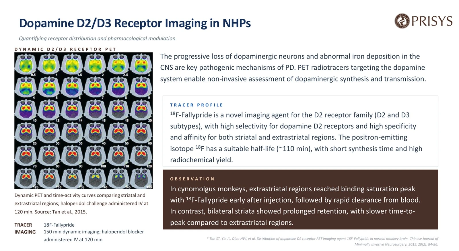
Prisys presented a working example during the Prisys Biotech webinar on molecular imaging in non-human primate (NHP) research. The study used [18F]Fallypride to follow dopamine D2/D3 receptor binding in cynomolgus monkeys through a 150-minute dynamic PET acquisition, with an intravenous haloperidol challenge at 120 minutes.
The case shows how dynamic PET characterizes regional receptor binding, demonstrates pharmacological displacement, and supports target engagement assessment in preclinical CNS programs.
1. Why Receptor Occupancy Matters Beyond Conventional PK
Receptor occupancy is the proportion of available target receptors bound by a drug at a given time. Plasma concentration and total tissue exposure describe how much drug is present. Occupancy describes what the drug is doing at the receptor.
For dopamine D2 antagonists and partial agonists, the relationship between plasma concentration and occupancy is nonlinear and saturable. At low concentrations, a modest increase in exposure produces a large increase in occupancy. As binding approaches saturation, further increases in concentration produce progressively smaller gains.
That shape has direct consequences for dose selection. Clinical PET studies of antipsychotic drugs have established associations between D2 receptor occupancy, therapeutic response, and extrapyramidal symptoms. Measuring occupancy therefore shows whether a candidate reaches an appropriate level of engagement without pushing exposure higher than the pharmacology requires.
The same logic applies to other dopamine-directed mechanisms. A D2 partial agonist needs enough occupancy to produce its effect while leaving room for endogenous dopaminergic signaling. Compounds aimed at extrastriatal receptors need demonstrable binding in those regions, and high striatal uptake alone does not show it.
2. What Does [18F]Fallypride PET Measure?
[18F]Fallypride is a substituted benzamide radioligand with high affinity for dopamine D2-like receptors, including the D2 and D3 subtypes. Binding is reversible, so the same tracer can be followed through association and dissociation within one session.
Its affinity is high enough to report binding in regions of low receptor density, including cortical and thalamic areas. Most other dopamine receptor tracers were developed for high-density regions and return little usable signal outside the striatum.
Fluorine-18 has a physical half-life of about 110 minutes, long enough for a dynamic protocol that captures delivery, regional accumulation, and washout in one acquisition.
Two interpretive limits apply. [18F]Fallypride does not separate D2 from D3, so the signal is D2/D3 binding and nothing more specific. PET signal intensity also does not equal receptor number. Measured radioactivity combines tracer delivery, nonspecific distribution, specific binding, and tracer kinetics, and separating those components requires kinetic analysis.
3. The NHP Case: Dynamic PET Imaging with a Haloperidol Challenge
Dopamine D2/D3 Receptor PET: [18F]Fallypride and the Haloperidol Challenge
Read receptor availability in the same animal, before and after a pharmacological challenge.
In this case study, Prisys presents dynamic [18F]Fallypride PET in cynomolgus monkeys, using a 150-minute acquisition with an intravenous haloperidol challenge at 120 minutes.
Why [18F]Fallypride, and how the study was designed:
- Binds dopamine D2-like receptors, so regional signal tracks receptor availability in the striatum and in lower-density extrastriatal areas such as cortex and thalamus
- Fluorine-18 labeling (about 110-minute half-life) supports a 150-minute dynamic acquisition in one session
- Haloperidol given at 120 minutes as a competing D2 antagonist, so displacement appears inside the same scan
- Striatal and extrastriatal regions compared for time to peak and for tracer retention
Keeping the animal in the same scanner takes inter-subject variability out of the comparison, and the challenge separates specific receptor binding from the rest of the signal. The scan is not a receptor count; it measures ligand binding, and binding potential comes out of the kinetic model.
Speaker: Dr. Wei Liu, Prisys Biotechnologies. Case from the Prisys Biotechnologies molecular imaging platform.
Please contact: bd@prisysbiotech.com.
Dr. Liu's example used [18F]Fallypride PET in cynomolgus monkeys to characterize tracer distribution and regional receptor binding in the normal brain. The aim was a baseline description rather than a disease model or a therapeutic candidate.
The acquisition ran for 150 minutes. At 120 minutes, haloperidol was given intravenously as a pharmacological blocking challenge.
Striatal and extrastriatal regions behaved differently. Extrastriatal regions generally peaked earlier and cleared faster. The bilateral striata reached peak later and retained tracer longer.
Receptor density explains the pattern. In a high-density region such as the striatum, most of the signal comes from specific binding. Where density is lower, tracer delivery and nonspecific distribution carry proportionally more of it.
That difference constrains study design. A single static image at one fixed time point cannot characterize striatal and extrastriatal binding at once, so acquisition timing and the analysis plan have to account for regional kinetics.
The haloperidol challenge added a pharmacological test. Haloperidol is a dopamine receptor antagonist and competes with [18F]Fallypride for the same binding sites. After dosing, tracer binding falls in the regions where the signal is largely specific D2/D3 binding. That displacement supports both the specificity and the reversibility of the regional signal.
4. How Are Receptor Occupancy and Binding Potential Quantified?
Images alone cannot carry a quantitative pharmacological claim. Most of the information sits in the time-activity curve (TAC), which traces radioactivity concentration in a selected region across the acquisition. Its shape reflects tracer delivery, receptor association, dissociation, and clearance.
For reversible tracers, quantification usually relies on a reference region with negligible specific binding to the target. That region estimates nondisplaceable tracer distribution and lets the analysis separate specific binding from the rest of the signal.
The parameter most often reported is the nondisplaceable binding potential (BPND), the ratio of specifically bound tracer to nondisplaceable tracer at equilibrium under the assumptions of the chosen kinetic model. Reference-tissue models or other validated kinetic approaches estimate it, depending on tracer and acquisition design.
Occupancy then follows from a comparison of binding potential before and after a competing drug:
Receptor Occupancy (%) = [(BPND, baseline − BPND, drug) / BPND, baseline] × 100
A fall in BPND after dosing means fewer sites remain available to the radioligand. Under the study assumptions, that fall estimates receptor occupancy.
The arithmetic is simple. The reliability is not. Physiological stability, consistent acquisition, accurate region definition, motion correction, and an appropriate kinetic model all feed into the estimate.
The challenge design in the webinar applies the same principle inside one session. Instead of comparing separate baseline and post-treatment scans, the blocking agent is given during a single dynamic acquisition, and displacement is read directly from the time-activity curve. Fewer imaging sessions means less between-session variability to control. It also puts weight on the design choices: challenge timing, blocking dose, and the length of acquisition remaining after dosing all determine whether displacement is captured well enough to measure.
5. What Can a Single-Dose Challenge Demonstrate?
A single-dose challenge tests tracer specificity and reversible target binding. It does not produce a dose-occupancy relationship.
That limit matters when PET results feed a development decision. One blocking dose is one intervention point. Displacement at that point supports a target-mediated reading of the signal, and says nothing about how occupancy moves across concentrations.
A quantitative dose-occupancy relationship needs several dose levels, ideally with plasma PK measured alongside so that occupancy can be related to systemic exposure. Such a design maps concentration against engagement, identifies the exposure range tied to a given occupancy band, and supports pharmacological dose selection.
Three further points bear on interpretation.
Pharmacological selectivity: haloperidol is not selective for D2/D3. It also interacts with sigma-1 and serotonin 5-HT2A receptors. Its use as a blocking agent is therefore read against the regional tracer distribution and the observed displacement, not as evidence that every effect runs through D2/D3.
Physiological conditions: anesthesia and physiological change affect cerebral blood flow, tracer delivery, and possibly endogenous dopaminergic activity. These variables need control and documentation across the imaging session.
Occupancy versus efficacy: occupancy shows target engagement, not therapeutic benefit. A compound can occupy its receptor substantially without producing the intended downstream response.
Receptor imaging therefore belongs alongside other pharmacodynamic endpoints rather than standing in for them.
6. Why Use Non-Human Primates for Dopamine Receptor PET Studies?
NHP imaging suits quantitative CNS receptor work, especially when the question covers striatal and extrastriatal populations together.
Rodent PET is well established, but the small brain limits regional quantification. Partial volume effects grow serious once a region of interest approaches the spatial resolution of the scanner, and separating adjacent structures becomes unreliable. Small extrastriatal regions suffer most.
A larger brain allows practical delineation of the striatum, thalamus, and cortical areas. NHPs also bridge rodent pharmacology and clinical neuroimaging: neuroanatomical organization and the dopaminergic system resemble the human arrangement closely enough to make distribution, receptor binding, and pharmacological response measurable in a translational setting.
Repeated imaging in the same animal is the other advantage. Within-subject designs reduce the influence of inter-individual variation in receptor availability and physiology, which matters when the expected effect is small or when binding has to be tracked over time.
NHP data are not interchangeable with human PET measurements. Physiology, tracer kinetics, anesthesia, and experimental conditions differ, and those differences have to be handled when quantitative findings are translated. Matched imaging modalities and analytical methods narrow the methodological gap.
7. Integrating Receptor Imaging into CNS Drug Development
Receptor PET contributes most when it sits inside a broader pharmacology and PK/PD strategy.
Target engagement assessment comes first. Before committing to larger efficacy studies, a team can establish whether a candidate reaches the brain and occupies its receptor at relevant systemic exposures.
Dose selection follows. Occupancy measured across several dose levels and integrated with plasma PK gives an exposure-occupancy relationship that provides a mechanistic basis for choosing doses in later studies, early clinical work included.
Candidate comparison is the third use. Where several compounds share a molecular target, PET occupancy data complement in vitro binding affinity and conventional PK, and allow in vivo engagement to be compared at matched exposures.
Read alongside other endpoints, these measurements describe the program more completely.
|
Measurement
|
Primary question addressed
|
|
Plasma PK
|
How much drug is present in systemic circulation?
|
|
CSF concentration
|
What is the measured drug concentration in CSF?
|
|
Biodistribution imaging
|
Where does the compound or radiolabeled material distribute?
|
|
Receptor PET
|
How much of the target is available for radioligand binding, and how does this change following drug administration?
|
|
Functional and behavioral endpoints
|
Does target engagement translate into measurable pharmacological effects?
|
Combining these measurements connects exposure, target engagement, and downstream pharmacological response.
8. Practical Considerations and Limitations of NHP Receptor PET
Quantitative receptor PET rewards careful design and tracer-specific validation.
The reference-region assumption underpins most quantitative approaches. The chosen region needs negligible specific binding to the target, and its suitability has to be established for the tracer and species in use.
Motion matters during long dynamic acquisitions. Small movements shift anatomical registration and distort regional time-activity curves, which propagates into the binding estimates.
Test-retest variability needs characterization whenever a change in binding is a primary endpoint. Without a baseline variability figure, a genuine pharmacological effect and a measurement difference look the same.
Tracer binding characteristics and off-target interactions should be checked in advance. Where subtype-specific information is required, [18F]Fallypride cannot separate D2 from D3 contributions.
These requirements set the conditions for using receptor PET: an appropriate tracer, defined quantitative endpoints, and analysis criteria agreed before the study starts.
Conclusion
[18F]Fallypride PET in cynomolgus monkeys shows what receptor imaging adds in vivo: regional D2/D3 binding, striatal and extrastriatal kinetics followed across a 150-minute dynamic acquisition, and pharmacological displacement after an intravenous haloperidol challenge at 120 minutes.
The wider contribution is the question it answers. Receptor PET establishes whether a candidate engages its intended target at a given exposure, which no plasma PK curve, CSF sample, or biodistribution image resolves on its own.
Read with conventional PK, CSF sampling, biodistribution imaging, and functional pharmacodynamic endpoints, occupancy data complete the chain from exposure to pharmacological activity.
Exposure, target engagement, and therapeutic efficacy stay distinct quantities. Keeping them distinct is what makes a translational study informative and a development decision defensible.
The Molecular Imaging Webinar Case Series
This article closes the six-part Prisys Biotech webinar series on molecular imaging in NHP research. Each part took one question.
The first followed whole-body biodistribution imaging in NHPs with iodine-125 SPECT, asking how a labeled compound distributes across organs over time. The second asked whether an intranasally administered compound reaches the brain, and used nose-to-brain delivery measured by SPECT to answer it. The third worked through Actinium-225 human dose projection from NHP imaging data, including absorbed-dose estimation and human activity estimates. The fourth tracked longitudinal tau PET in a rhesus macaque model. The fifth applied VMAT2 PET in a non-human primate model of Parkinson's disease to quantify a dopaminergic deficit in vivo.
|
No.
|
Topic
|
Key Research Question
|
|
1
|
How does a radiolabeled compound distribute across organs over time?
|
|
|
2
|
Does an intranasally administered compound reach the brain, and how long is it retained?
|
|
|
3
|
How can preclinical imaging data support absorbed-dose estimation and human activity projection?
|
|
|
4
|
How does tau-associated imaging signal change over time?
|
|
|
5
|
Can dopaminergic deficits associated with Parkinsonian pathology be quantified in vivo?
|
|
|
6
|
How can PET imaging evaluate receptor availability and pharmacological target engagement?
|
Read as a set, the six move from distribution through delivery, dosimetry, disease progression, and dopaminergic deficit to receptor availability, which is the point at which an imaging readout starts to answer a pharmacology question.
Selecting an imaging method follows the scientific question. Different tracers, acquisition protocols, and analytical approaches return complementary information, and the combination strengthens interpretation of preclinical pharmacology data.
For further context, the series is accompanied by an overview article on molecular imaging applications in NHP research and translational drug development.
Frequently asked questions
Q: What is dopamine D2/D3 receptor occupancy in PET imaging?
A: Dopamine D2/D3 receptor occupancy is the proportion of available receptors bound by a drug. PET imaging with a reversible radioligand such as [18F]Fallypride quantifies the change in ligand binding after a pharmacological intervention, which gives an in vivo measure of target engagement.
Q: Why is [18F]Fallypride used for dopamine receptor imaging?
A: [18F]Fallypride has high affinity for D2-like receptors and reports binding in both striatal and extrastriatal brain regions. Reversible binding makes it suitable for dynamic PET studies and pharmacological displacement experiments.
Q: What is the purpose of a haloperidol challenge during [18F]Fallypride PET?
A: Haloperidol is a competing dopamine receptor antagonist. Given during a dynamic PET acquisition, it lets researchers observe the change in [18F]Fallypride binding. A reduction in regional tracer binding supports the reading that the signal includes specific, reversible D2/D3 receptor binding.
Q: Can a single-dose PET challenge establish a dose-occupancy relationship?
A: No. A single-dose challenge reports pharmacological displacement at one intervention point. A dose-occupancy relationship requires several dose levels, ideally with plasma PK measurements that relate systemic exposure to receptor occupancy.
Q: What is the difference between receptor occupancy and drug efficacy?
A: Receptor occupancy measures target engagement. Drug efficacy refers to the ability of a compound to produce its intended pharmacological or therapeutic effect. High occupancy does not necessarily translate into a beneficial functional response, so both measurements belong in the evaluation of a CNS candidate.
Q: Why are non-human primates useful for CNS receptor PET studies?
A: NHPs offer a larger brain for regional PET analysis and close neuroanatomical and physiological similarity to humans. Repeated imaging in the same animal also allows longitudinal assessment of receptor binding and pharmacological response.