For a conventional small-molecule or antibody program, the starting dose in a first-in-human (FIH) study is usually considered in relation to mass dose, systemic exposure, pharmacology, and toxicology. Radiopharmaceutical development adds another variable: the administered activity determines how much radiation is delivered to the tumor and to healthy tissues.
For an actinium-225 (Ac-225) therapy, the relevant question is therefore not only how much compound reaches the circulation. Investigators also need to estimate where the radiolabeled material accumulates, how long it remains in each organ, and what absorbed radiation dose may result. Tumor-to-organ dose relationships are an important part of this assessment, but they are considered together with pharmacology, toxicity, manufacturing, and other nonclinical evidence.
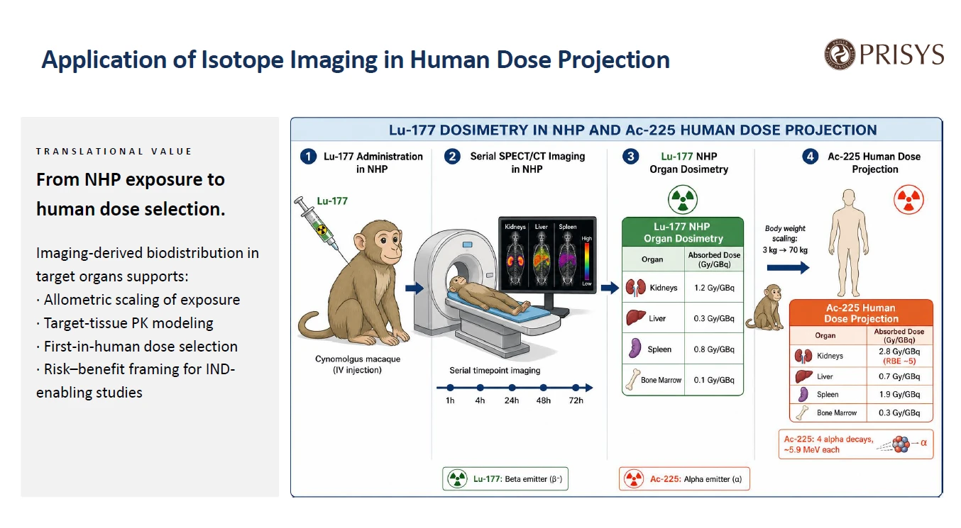
Preclinical molecular imaging can provide part of this evidence. A targeting vector can be labeled with a suitable imaging radionuclide and followed by serial PET or SPECT imaging in non-human primates (NHPs). The resulting organ-level time-activity data can be used to estimate residence times and support dosimetry calculations. These data may then be scaled to a reference human model to inform an initial Ac-225 activity estimate.
This article is the third in a series based on the Prisys Biotech molecular imaging webinar. The previous article described whole-body biodistribution in NHPs using serial imaging and %ID/g measurements. Those distribution data can provide an input to a dosimetry workflow, provided that the tracer, labeling chemistry, image quantification, and therapeutic construct are sufficiently comparable for the intended analysis.
Why radiopharmaceutical dosing requires more than plasma PK
In a conventional development program, exposure-based safety margins are often derived from plasma PK parameters such as area under the curve (AUC), maximum concentration, and clearance, together with toxicology findings. Radiopharmaceuticals require an additional assessment of tissue-associated activity and radiation dose.
Ac-225 is an alpha-emitting radionuclide with a physical half-life of approximately 10 days. Alpha particles have a short tissue range, generally on the order of several cell diameters, and deposit their energy close to the site of decay. As a result, the biological effect depends strongly on the location and residence time of the radionuclide. The organs that limit activity may include the kidneys, liver, bone marrow, or salivary glands, depending on the targeting vector, chelator, formulation, administration route, and biological disposition.
Before a patient is treated, the development team needs an estimate of activity distribution over time in the organs relevant to efficacy and safety. In practical terms, this requires time-activity curves for the target tissue and organs at risk. Terminal tissue studies can provide direct measurements, but each animal generally contributes data from one scheduled time point. A full time course therefore requires several terminal groups, and the measurements are affected by inter-animal variability.
Large-animal imaging adds a longitudinal in vivo component. It does not remove the need for direct tissue measurements, but it can show how organ-associated activity changes over time in the same subject.
How imaging-based dosimetry is performed
The general approach is to use an imaging radionuclide on the same, or a closely matched, targeting vector that will carry the therapeutic radionuclide. The labeled test article is administered, often at tracer mass in a microdose design, and the animals undergo serial PET or SPECT imaging.
The choice of imaging modality and radionuclide depends on the biological kinetics of the vector. A short-lived isotope may be suitable for a small molecule with rapid distribution and clearance. A longer-lived isotope may be required for an antibody or another vector with slower tissue uptake and retention.
Serial images are processed to estimate activity in predefined organs or regions of interest. The resulting organ-level activity data are plotted over time and integrated to estimate residence time. The residence time is then combined with the physical decay characteristics and emission data of the intended therapeutic radionuclide, such as Ac-225, within an absorbed-dose model.
The NHP estimates can subsequently be scaled to a reference human model. This step takes account of differences in body mass, organ mass, and, depending on the model, other anatomical parameters. The output is an estimated absorbed dose per unit of administered activity for the organs included in the model. It can be used to compare target-tissue exposure with exposure in organs that may limit treatment activity.
The calculation is not independent of the biological assumptions. It depends on the quality of the image data, organ segmentation, calibration, the selected kinetic model, the relationship between the imaging tracer and the therapeutic construct, and the treatment of uncertainty during interspecies scaling.
Case study: serial NHP SPECT and Ac-225 dose projection
Human Dose Projection and Organ Dosimetry: Serial SPECT Imaging in NHPs With Ac-225
How can serial molecular imaging in non-human primates support organ dosimetry and the projection of an initial Ac-225 activity?
In this case study from the Prisys Biotech webinar, Prisys describes how serial SPECT imaging can follow the distribution of a radiolabeled test article over time and provide organ-level data for residence-time estimation and absorbed-dose modeling.
Study highlights:
- Radiolabeled test article administered intravenously
- Serial SPECT imaging used to monitor whole-body distribution
- Organ-level time-activity data used to estimate residence time
- NHP data scaled to a reference human model for Ac-225 activity projection
Imaging-based dosimetry can connect biodistribution data with organ-level absorbed-dose estimates. The resulting projection remains dependent on the imaging surrogate, labeling stability, dosimetry model, interspecies scaling, and complementary nonclinical and clinical evidence.
Case from the Prisys Biotech Molecular Imaging platform.
Please contact: bd@prisysbiotech.com.

The webinar described Prisys Biotech's NHP translational research capabilities and radiochemistry and molecular imaging capabilities.
In the case presented, a radiolabeled test article was administered intravenously to NHPs. Serial SPECT imaging was then used to follow whole-body distribution over time. The organ-level activity data were analyzed to support a radionuclide dosimetry calculation, and the NHP results were scaled to a reference human body-weight model for an Ac-225 activity projection.
The same imaging dataset can support two related analyses. The first is a biodistribution assessment, which describes organ-associated signal and may include measurements such as %ID/g. The second is a dosimetry analysis, which uses the time course of activity to estimate residence time and absorbed dose. These analyses are related but not interchangeable. A biodistribution result is not automatically a validated absorbed-dose estimate, and a dosimetry result remains dependent on the assumptions used in the calculation.
A longitudinal design also avoids treating each time point as an entirely separate observation. Repeated imaging in the same animal can reduce the contribution of between-subject variation to the observed time course. Whether it reduces the number of terminal animals depends on the study objectives and on the amount of ex vivo confirmation required.
Selecting an imaging surrogate
Radionuclide selection should follow the expected biological time course and the chemistry of the targeting vector. The study team needs to consider two separate half-lives:
- the physical half-life of the imaging radionuclide; and
- the biological half-life of the labeled molecule in blood and tissues.
The imaging isotope must remain detectable for long enough to capture the relevant distribution and clearance phases. For example, gallium-68 has a physical half-life of about 68 minutes. That may be appropriate for some rapidly distributed molecules, but it would not normally capture the later kinetics of an antibody that circulates for days or weeks. Longer-lived radionuclides, such as zirconium-89 for PET or iodine-125 for SPECT, may be considered for slower biological processes when the labeling chemistry and study design are suitable. Fluorine-18 may be appropriate for compounds with faster kinetics.
The physical half-life is only one part of the decision. The label must remain associated with the vector for the period being modeled, and the labeling procedure should not materially alter the vector's distribution or biological activity. Specific activity, radiochemical purity, formulation, stability, and the relationship between the imaging construct and the therapeutic construct all require assessment.
A surrogate label does not automatically reproduce the behavior of an Ac-225-labeled construct. Differences in radionuclide, chelator, specific activity, molecular integrity, or formulation may change distribution. The projection should therefore be based on a documented comparability rationale rather than on isotope substitution alone.
What the study can contribute
An imaging-based dosimetry study may provide:
- organ-level time-activity data for the target tissue and relevant organs;
- estimates of residence time and absorbed dose per unit of administered activity;
- a comparison of target-tissue exposure with activity in organs that may limit treatment;
- biodistribution information from the same serial imaging dataset; and
- information that can be considered alongside PK, toxicology, pharmacology, and manufacturing data when an FIH activity is evaluated.
The value of these outputs depends on the study design and validation. They should be described as estimates or projections, not as direct measurements of the future patient dose.
What the projection cannot establish on its own
Several sources of uncertainty need to remain visible in the interpretation.
First, the projection depends on the assumption that the imaging tracer reports the distribution of the therapeutic construct. A difference in label stability, chelation, formulation, or molecular integrity can introduce a mismatch between the imaged material and the future Ac-225 product.
Second, Ac-225 decays through a chain of daughter radionuclides. Nuclear recoil during alpha decay can affect retention of daughter products in the chelator and may lead to redistribution of some daughters after decay. Imaging the parent vector does not fully characterize the distribution of every daughter isotope. Daughter redistribution and retention therefore require separate consideration in the dosimetry model and in the overall safety assessment.
Third, organ-level imaging cannot resolve the micrometre-scale distribution of alpha-particle energy within tissue. Two regions within the same organ may receive different cellular doses even when their average organ activity appears similar. Microdosimetry and cellular localization require methods beyond routine PET or SPECT.
Fourth, interspecies scaling is a model-based step. NHP anatomy and physiology may provide useful context for some programs, but the NHP-to-human relationship is not exact. Organ masses, receptor expression, metabolism, renal handling, and immunogenicity can all influence clinical disposition.
For these reasons, a preclinical dose projection should be treated as an input to FIH planning. Clinical imaging and pharmacokinetic measurements after initial administration are needed to assess how well the preclinical projection describes patient biodistribution and to refine subsequent activity decisions.
Why include NHPs in dosimetry studies?
Rodent studies remain important for early biodistribution, pharmacology, and toxicology work. NHPs may add information when the development question depends on larger-animal anatomy, vascular access, organ dimensions, receptor biology, or serial imaging under conditions closer to the planned clinical procedure.
NHP imaging can generate repeated observations from the same animal, which is useful when estimating the shape of an organ time-activity curve. It can also provide whole-body context for organs that are difficult to assess from plasma PK alone. These advantages do not make NHP data universally superior, and the species should be selected according to the target, vector, therapeutic mechanism, expected kinetics, and regulatory strategy.
Molecular imaging and dosimetry studies at Prisys Biotech
Prisys Biotech supports NHP molecular imaging studies that can include radiolabel selection, labeling, serial PET/CT or SPECT/CT, organ-level image analysis, and integration with PK, biomarker, pharmacology, and safety assessments. The webinar described radiochemistry, isotope labeling, post-labeling preparation, purification, and quality-control procedures.
The appropriate workflow depends on the test article and the development question. Before a study begins, the team should define the therapeutic construct, imaging surrogate, labeling chemistry, expected kinetic window, organs of interest, image-quantification method, ex vivo confirmation plan, and the model used for human scaling.
For an Ac-225 program, the purpose of the study is to build a traceable chain of evidence from radiolabeled vector distribution to residence time and absorbed-dose estimates. The final activity decision must then incorporate this information with the broader nonclinical and clinical dataset.
Conclusion
Serial molecular imaging in NHPs can support the development of an Ac-225 activity projection by providing organ-level distribution data over time. When the imaging tracer is sufficiently comparable to the therapeutic construct, these data can be used to estimate residence times and inform absorbed-dose calculations after scaling to a reference human model.
The approach has clear boundaries. It does not directly measure the future patient dose, fully characterize daughter radionuclide redistribution, resolve cellular alpha-particle dose, or eliminate uncertainty in interspecies scaling. It is most useful as one part of an integrated program that includes radiochemistry, image validation, PK, biodistribution, dosimetry modeling, toxicology, pharmacology, and early clinical imaging.
Frequently asked questions
Q: Why is a surrogate imaging isotope used instead of Ac-225 itself?
A: Ac-225 is not generally selected as the primary radionuclide for routine quantitative imaging of whole-body kinetics. A surrogate imaging isotope on the same or closely matched targeting vector can provide the time-activity data needed for the dosimetry model. The relationship between the surrogate and the therapeutic construct must be assessed rather than assumed.
Q: How are NHP imaging data scaled to humans?
A: Organ time-activity curves are integrated to estimate residence time. The therapeutic radionuclide's decay and emission characteristics are then applied in an absorbed-dose model. The resulting NHP estimates are scaled to a reference human model using parameters such as body mass and organ mass, together with the assumptions of the selected model.
Q: Which imaging isotope is appropriate for the surrogate?
A: The choice depends on the molecular kinetics, expected tissue-retention window, labeling chemistry, imaging modality, and study objectives. Shorter-lived isotopes may suit rapidly distributing small molecules, while longer-lived isotopes may be more appropriate for antibodies or other slowly distributing vectors. The isotope must also remain associated with the vector for the period being modeled.
Q: Can one imaging study support both biodistribution and dosimetry?
A: It can, if the acquisition schedule, calibration, image analysis, and data quality are suitable for both purposes. The same serial images may provide organ-associated activity data for biodistribution and the time course needed for residence-time calculations. These are separate analyses and should be validated separately.
Q: What are the main uncertainties in an Ac-225 dose projection?
A: Important uncertainties include the comparability of the imaging surrogate and therapeutic construct, label stability, daughter-radionuclide redistribution, organ segmentation and calibration, the dosimetry model, and interspecies scaling. Early clinical imaging and PK are needed to evaluate the projection in patients.