Parkinson's disease (PD) is defined clinically by its motor features, while the biology underneath is the loss of dopaminergic neurons in the nigrostriatal system. For a therapeutic program, symptom improvement in an animal answers part of the question. The sponsor also needs to know whether the drug acts on that disease biology, and whether the effect can be measured while the study is running.
Molecular imaging is one route to that question. A radioligand that binds a specific molecular target lets PET quantify dopaminergic function in the brain of a living animal, which behavioral scores cannot do on their own. In webinar on molecular imaging in non-human primate (NHP) research, Prisys described an NHP study that used [18F]AV-133, a ligand for vesicular monoamine transporter 2 (VMAT2), to assess dopaminergic integrity in a cynomolgus monkey model of PD. The design combined a unilateral lesion with serial PET/CT and PET/MRI, so tracer distribution could be compared before and after treatment.
Why dopaminergic imaging matters in PD
VMAT2 packages dopamine into synaptic vesicles. The transporter is expressed in monoaminergic neurons, and its density in the striatum tracks the integrity of the nigrostriatal system. In PD, progressive loss of dopaminergic neurons in the substantia nigra reduces dopamine storage and lowers striatal VMAT2 signal.
Conventional assessment of a PD model often relies on motor behavior, a downstream readout of neuronal function. Behavior is informative, and it is also influenced by factors outside the target pathology: training, motivation, sedation, and compensation by other neural circuits. A molecular imaging endpoint answers a different question. It measures the expression or function of a protein target in a defined brain region, in the same animal, across time points.
For a therapeutic candidate, an imaging biomarker addresses three questions. Has the drug engaged the intended system? Is the response anatomically consistent with the pathology? Does the change hold across repeated measurements? Imaging does not establish that a treatment reversed neuronal loss. It does support a more specific interpretation than behavior alone.
How VMAT2 PET imaging works
A PET study with [18F]AV-133 begins with a trace dose of the radiolabeled ligand, given intravenously. Fluorine-18 has a physical half-life of about 110 minutes, long enough for a single imaging session. The ligand crosses the blood-brain barrier and binds VMAT2 in the brain. The scanner maps the distribution of the radioactive signal, and image analysis then quantifies uptake in defined brain regions.
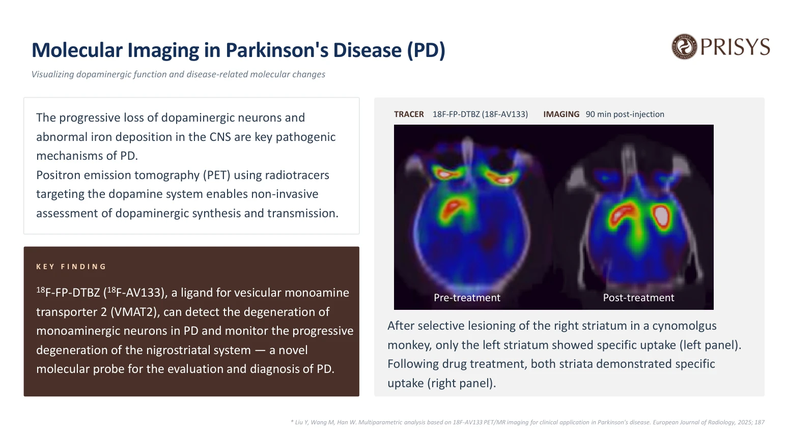
In the webinar case, scans were acquired 90 minutes after tracer administration, on both PET/CT and PET/MRI, which gives the molecular signal co-registered anatomical context. Because the ligand binds VMAT2, regional signal is read as a marker of VMAT2 expression or density in the imaged area.
Several factors shape that reading: the specific binding of the tracer, the interval between injection and scan, the animal's physiological state, image reconstruction and registration, and how the region of interest is drawn. A difference between two scans therefore has to be interpreted against the study design and confirmed with appropriate controls.
Case study: VMAT2 imaging in a lesioned cynomolgus monkey model
NHP Parkinson's Disease Imaging: VMAT2 & Dopamine D2 PET Tracers
Read dopaminergic integrity in the same animal, before and after treatment.
In this case study, Prisys presents longitudinal VMAT2 PET in a cynomolgus monkey model of Parkinson's disease, using [18F]AV-133 in a unilateral striatal lesion model.
Why [18F]AV-133, and how the study was designed:
- Binds vesicular monoamine transporter 2 (VMAT2), so regional signal tracks the density and function of monoaminergic terminals
- Fluorine-18 labeling (about 110-minute half-life) supports a single imaging session after intravenous injection
- Selective lesion of the right striatum, creating a unilateral dopaminergic deficit
- Serial PET/CT and PET/MRI at 90 minutes after tracer administration, before and after treatment
Imaging the same animal before and after treatment takes inter-subject variability out of the comparison, and co-registered MRI anchors the signal to anatomy. The scan is not a neuron count; it measures ligand binding to VMAT2.
Speaker: Prisys Biotechnologies. Case from the Prisys Biotechnologies molecular imaging platform.
Please contact: bd@prisysbiotech.com.
The webinar described a study in a cynomolgus monkey with a selective lesion of the right striatum, created to model a dopaminergic deficit. Before treatment, [18F]AV-133 PET showed specific tracer uptake in the left striatum and reduced signal on the right, an asymmetry consistent with the induced unilateral lesion.
After treatment with a test article, a follow-up scan showed specific uptake returning in the right striatum. The presentation described this as recovery of VMAT2-associated signal on the previously lesioned side. Both scans were acquired 90 minutes after tracer administration, on PET/CT and PET/MRI.
The case shows a targeted radioligand giving an in vivo readout of dopaminergic integrity in a defined brain region. The comparison was made within one animal, which removes inter-subject variability from it. Co-registered MRI anchors the signal to anatomy and sharpens the regional analysis.
The study does not measure neuronal number, and it does not establish that treatment reversed the underlying pathology. The change in tracer signal is compatible with a treatment-related effect on VMAT2 expression or function. Altered tracer delivery, changed vesicular packaging, and recovery of presynaptic function without restoration of neuronal count would produce a similar image. Complementary endpoints are needed to separate those explanations.
What the imaging can, and cannot, show
A VMAT2 ligand shows regional differences in tracer binding at dopaminergic terminals. In a unilateral lesion model, that means showing whether the affected side differs from the intact side, and whether treatment changes the size of that difference. The longitudinal design keeps the comparison within one animal across time points.
PET signal is not a count of dopaminergic neurons. It reflects ligand binding to VMAT2, which depends on the number of terminals, the expression level of the transporter, and the functional state of the vesicular system. A change in signal does not separate neuronal survival from axonal sprouting, altered vesicle turnover, or other mechanisms that shift VMAT2 availability.
Reading an imaging result as disease modification requires behavioral assessment, histological analysis of dopaminergic markers, and other measures of neuronal integrity alongside it. The webinar case shows how molecular imaging fits into that integrated evaluation. It does not replace tissue-level confirmation.
Why NHPs for PD imaging studies
Rodent models of Parkinson's disease are widely used for early screening and mechanistic work, and their brain size and neuroanatomy limit imaging resolution and regional specificity. An NHP brain is larger, with a basal ganglia organization closer to the human one, and can be imaged on clinical-equivalent systems and protocols. That matters when the study question involves regional tracer distribution, quantitative image analysis, or a direct read-across to human imaging endpoints.
Species choice follows the scientific question. Where a program needs regional assessment of dopaminergic integrity, repeated imaging, or integration with surgery or a delivery procedure, an NHP model offers more than a rodent. The model still has to be validated for the specific tracer and disease mechanism in play.
Integrating imaging with other endpoints
Imaging is more useful in a PD study when complementary measures run alongside it. Behavioral assessment gives functional evidence of motor impairment and recovery. Histology and immunohistochemistry confirm the survival and morphology of dopaminergic neurons. Fluid biomarkers such as dopamine metabolites or alpha-synuclein species add information on disease state.
Plan the imaging endpoint together with those measures. Tracer choice, scan timing, region definition, and quantification method stay consistent across animals and time points. When imaging supports a therapeutic claim, the design needs controls and a pre-specified analysis plan, so that a single scan is not over-read.
Molecular imaging in PD research at Prisys Biotech
Prisys Biotech's webinar presented VMAT2 imaging as one application of its NHP molecular imaging platform. The case paired a unilateral striatal lesion with serial PET/CT and PET/MRI, using [18F]AV-133 to assess dopaminergic integrity before and after treatment. Tracer selection, scan timing, image quantification, and the link to behavioral or histological endpoints are decided per study, against the biological question and a validated protocol.
The webinar also covered other molecular imaging applications in NHPs: tau PET, intranasal CNS delivery, and whole-body biodistribution. Each uses a different tracer and answers a distinct question, so their designs are not interchangeable.
Conclusion
Serial PET with a VMAT2 ligand measures dopaminergic integrity in vivo in an NHP model of PD. In the webinar case, a cynomolgus monkey with a unilateral striatal lesion showed asymmetric tracer uptake before treatment and a return of signal on the lesioned side afterwards. Molecular imaging tracked the disease-related change and gave the candidate a measurable readout.
Read the result against tracer specificity, scan timing, image analysis, and the endpoints run alongside it. A change in VMAT2-associated signal is consistent with a treatment effect on dopaminergic function. On its own it does not establish neuronal recovery or disease modification. A translational interpretation needs behavior, histology, and other biomarkers in the same design.
Reference
Liu Y, Wang M, Han W. Multiparametric analysis based on 18F-AV133 PET/MR imaging for clinical application in Parkinson's disease. European Journal of Radiology. 2025;187. The webinar slide cited this paper for tracer and clinical context. It does not describe the webinar study design or results.
Frequently asked questions
Q: What does VMAT2 PET measure in a PD model?
A: VMAT2 PET maps a radiolabeled ligand that binds vesicular monoamine transporter 2. The signal tracks the density and function of monoaminergic terminals in the imaged brain region. It is not a count of dopaminergic neurons.
Q: What was the design of the webinar case?
A: A cynomolgus monkey with a selective lesion of the right striatum. [18F]AV-133 PET/CT and PET/MRI were acquired 90 minutes after tracer administration, before and after treatment with a test article. The pre-treatment scan showed asymmetric uptake; the post-treatment scan showed signal returning on the lesioned side.
Q: Can the imaging result be read as evidence of therapeutic efficacy?
A: Returning tracer signal is consistent with a treatment-related effect on VMAT2-associated binding. It does not establish that treatment reversed neuronal loss or improved clinical function. The result needs behavioral, histological, and other measures behind it before it supports a broader efficacy claim.
Q: Why use NHPs for this type of study?
A: An NHP brain is larger than a rodent brain and its basal ganglia organization is closer to the human one, which supports regional imaging and quantitative analysis. Imaging can run on clinical-equivalent systems, which improves the translational relevance of the endpoint.
Q: What are the main limitations of VMAT2 PET in PD research?
A: The signal reflects ligand binding, not neuronal number. Tracer delivery, changes in expression, and functional state all shift it. Regional analysis depends on image registration and region definition. Confirm the result with tissue-level and functional measures.