Non-invasive imaging adds a longitudinal dimension to non-human primate (NHP) liver studies, but the different MRI techniques measure different biological features. MRI-derived proton density fat fraction (MRI-PDFF) estimates the fraction of mobile protons attributable to fat relative to the combined fat and water signal. It quantifies hepatic steatosis; it does not measure collagen or fibrosis directly. Liver biopsy provides tissue for histopathologic assessment, including fibrosis staging, but it samples only a small part of the organ.
For a fibrosis question, the imaging measure of interest is a fibrosis-oriented one, such as MRE-derived liver stiffness or MRI-derived extracellular volume (ECV), read alongside biopsy. PDFF complements those endpoints when steatosis is part of the model or the treatment hypothesis.
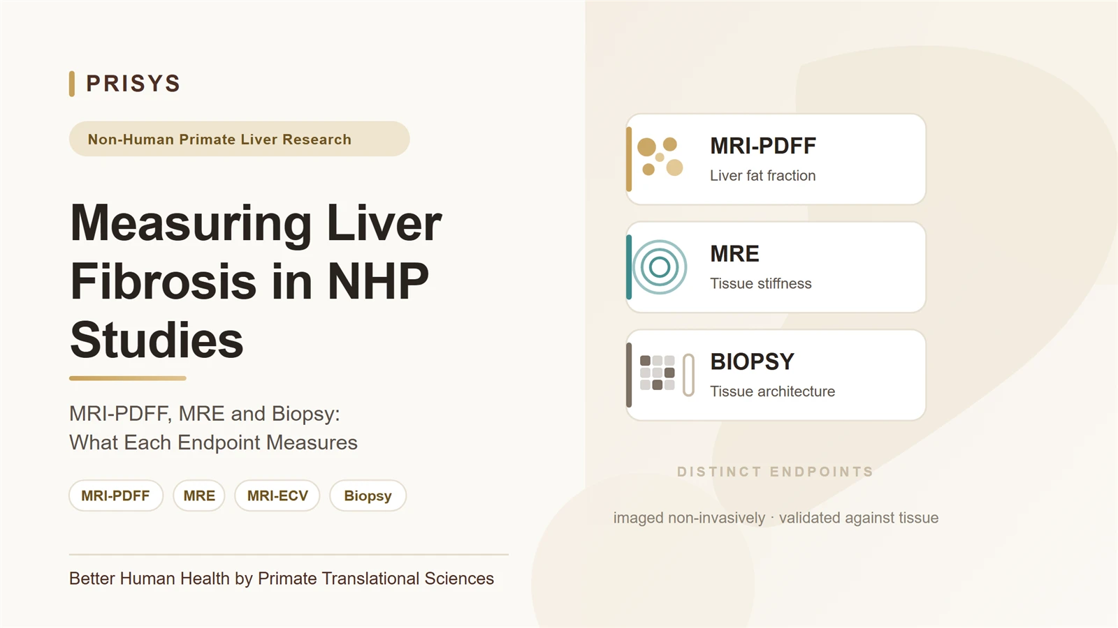
Match the imaging method to the endpoint
MRI-PDFF is a quantitative measure of liver fat. It is useful in diet-induced and other steatotic liver models when the study asks whether hepatic fat changes over time or in response to an intervention. A change in PDFF means a change in the measured fat fraction. On its own it is not evidence that fibrosis has regressed or progressed.
MRE estimates tissue stiffness from the propagation of mechanically induced shear waves. Liver stiffness is associated with fibrosis, but the measurement is indirect and other factors, including inflammation, congestion, cholestasis, acquisition conditions and technical quality, also affect it. MRI-derived ECV is a different parameter, generally calculated from pre- and post-contrast T1 measurements with hematocrit correction. The two methods are not interchangeable.
Biopsy allows direct examination of sampled tissue. Depending on the protocol, pathology reports fibrosis stage, steatosis, inflammation, hepatocyte ballooning, bile-duct changes and other features. Histology samples a small part of the organ, not the whole liver, and sampling location, specimen adequacy, staining, scoring method and reader variability all affect the result. In NHP studies, repeat sampling also has to be justified against animal welfare, procedural burden and the scientific value of the information.
The methods differ in what they measure and how the results should be interpreted:
- MRI-PDFF measures liver fat fraction. It quantifies steatosis and its change over time; it does not quantify fibrosis directly.
- MRE estimates shear-wave-derived tissue stiffness. It can support non-invasive fibrosis assessment where validated, but stiffness is indirect and non-fibrotic factors also affect it.
- MRI-ECV estimates contrast-enhanced extracellular volume fraction. Its use for fibrosis assessment is protocol-specific and requires validation, and it is not interchangeable with MRE.
- Biopsy with histology characterizes features in sampled liver tissue, including fibrosis stage and tissue injury. Sampling is limited, and scoring and repeatability require control.
What NHP studies show, and what they do not
Published NHP work provides examples of MRI-based liver measurements paired with histology, though the methods and models have to be named precisely. In a prospective study of 48 cynomolgus monkeys, liver ECV was measured in animals with non-alcoholic steatohepatitis and compared with histologic fibrosis staging. That study evaluated MRI-derived ECV; it is not validation of MRE or MRI-PDFF for fibrosis.[1]
A later report studied 104 cynomolgus monkeys with biopsy-confirmed early fibrosis (F1) and 10 healthy controls. It assessed MRI-ECV together with PDFF. That report linked ECV to early fibrosis assessment and PDFF to fat quantification; PDFF correlated with the histologic steatosis component of the NAFLD Activity Score (NAS). The results support using the two MRI measures for different endpoints in that cohort. They do not establish PDFF as a fibrosis measure, and they do not define transferable cut-offs for other species, disease models, scanners or laboratories.[2]
Human studies help explain the intended distinction between the modalities, but they are not a substitute for NHP validation. A longitudinal analysis in patients with NASH, for example, evaluated MRE-stiffness against fibrosis response and MRI-PDFF against steatosis response, using biopsy findings as the comparator. The authors described the results as preliminary and called for further evaluation.[3] Human performance estimates are not established NHP operating characteristics.
Biopsy remains informative, but is not a whole-liver reference
A biopsy distinguishes tissue features that imaging does not resolve, and it provides a fibrosis stage based on a prespecified scoring system. It also helps establish whether a model is characterized mainly by steatosis, steatohepatitis, cholestatic injury or another pathology. A core biopsy, however, represents a small tissue sample. A focal sample may not reflect the distribution of lesions across the liver, and apparent change between time points may partly reflect sampling or reader variation.
Scoring terminology also matters. In the commonly used NAFLD activity score framework, NAS is the unweighted sum of steatosis, lobular inflammation and hepatocellular ballooning. Fibrosis is assessed separately in the broader histologic scoring system and is not part of the NAS total.[4] If fibrosis is the primary endpoint, the protocol should state the fibrosis staging method and report it separately from NAS.
Study design considerations for serial NHP assessment
The model's etiology should guide the imaging package. A diet-induced steatotic or steatohepatitis model justifies measuring PDFF alongside a fibrosis-related endpoint. A bile-duct stenosis or ligation model represents cholestatic injury and should not be assumed to reproduce metabolic steatohepatitis. In either setting, a measure that works in one model will not necessarily work the same way in another.
When imaging and biopsy are used together, the protocol should define the primary endpoint before the study begins. Relevant details include the species and model, imaging sequence and scanner, acquisition timing, fasting and anesthesia conditions where applicable, regions of interest, image quality criteria, biopsy location and timing, histology stains, fibrosis scoring method, reader blinding, and the analysis plan. If repeated measurements are intended, within-animal repeatability and the expected biological range of change matter as well.
Paired imaging and histology characterize how an imaging measure relates to tissue findings in a particular model. That relationship has to be evaluated with prespecified methods and an adequate range of disease severity. Correlation alone does not establish that two endpoints are interchangeable, or that an imaging change predicts a treatment-related change in fibrosis.
Choosing endpoints by research question
If the primary question concerns liver fat, MRI-PDFF is an appropriate quantitative imaging endpoint, with histology or other study-specific measures providing complementary context. If the primary question concerns fibrosis, a fibrosis-oriented measure such as MRE or MRI-ECV applies only when the protocol and model support it; histologic fibrosis staging may be included as a tissue comparator. If the study concerns both steatosis and fibrosis, prespecify separate endpoints rather than combining them into a single interpretation.
In NHP liver research, the choice between imaging and biopsy depends on the question, not on which method is generally better. Imaging supports repeat, non-invasive measurement across the liver; biopsy characterizes sampled tissue. The value of either method depends on the biological endpoint, disease model, validation evidence and study design. MRI-PDFF is a liver-fat measure; fibrosis conclusions need fibrosis-specific evidence.
References
- Lyu L, Liu XL, Rui MP, et al. Liver extracellular volume fraction values obtained with magnetic resonance imaging can quantitatively stage liver fibrosis: a validation study in monkeys with nonalcoholic steatohepatitis. European Radiology. 2020. doi:10.1007/s00330-020-06902-w. PMID: 32377814.
- Rui MP, Zhu XY, Lü L, Liu XL, Fan D, Zhu ZH, et al. 磁共振细胞外容积分数联合PDFF成像在NAFLD食蟹猴模型中对早期肝脏纤维化的诊断价值 [Diagnostic value of MRI-ECV combined with MRI-PDFF imaging for early liver fibrosis in a NAFLD cynomolgus monkey model]. Chinese Journal of CT and MRI. 2024;(5):120–122. Journal record. (Chinese-language article.)
- Jayakumar S, Middleton MS, Lawitz EJ, et al. Longitudinal correlations between MRE, MRI-PDFF, and liver histology in patients with non-alcoholic steatohepatitis: Analysis of data from a phase II trial of selonsertib. Journal of Hepatology. 2019. doi:10.1016/j.jhep.2018.09.024. PMID: 30291868.
- Kleiner DE, Brunt EM, Van Natta M, et al. Design and validation of a histological scoring system for nonalcoholic fatty liver disease. Hepatology. 2005;41(6):1313–1321. doi:10.1002/hep.20701. PMID: 15915461.