Introduction: From Administered Dose to Target-Site Exposure
Dose selection in non-human primate (NHP) studies usually begins with a mg/kg conversion, and that conversion covers only the first step. The administered dose does not by itself predict systemic exposure, drug concentration at the target tissue, or the size of the pharmacological response.
NHP studies commonly bridge rodent and in vitro findings into clinical development. At that point the question set widens: whether a candidate produces a biological effect, what exposure produces it, how long that exposure must be maintained, and whether the drug reaches the intended site of action.
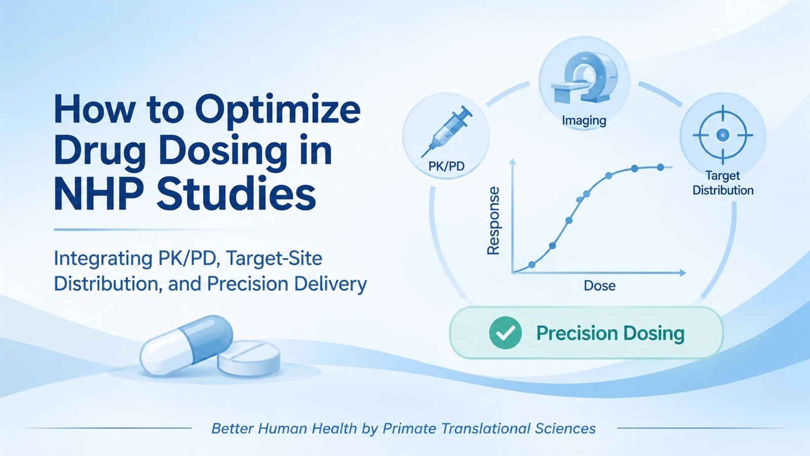
A workable NHP dose optimization strategy integrates four dimensions: pharmacokinetics (PK), pharmacodynamics (PD), target-site drug distribution, and experimental design. Together they give a basis for setting the starting dose, adjusting the dosing interval, and judging whether the observed response matches the intended mechanism of action.
1. Why Body-Weight-Based Dosing Is Not Enough
Body-weight-based dosing remains a practical starting point for many preclinical studies. Administering the same mg/kg dose to different animals still does not guarantee equivalent exposure.
Variability in absorption, distribution, metabolism, and excretion (ADME) drives most of that spread. Age, sex, physiological condition, disease status, administration route, formulation, and individual biological variation all contribute.
Which factors matter depends on the modality. Small molecules are cleared largely through hepatic metabolism and renal elimination. Monoclonal antibodies and other biologics circulate for weeks, show target-mediated drug disposition, and shift exposure when immunogenicity develops. Nucleic acid therapeutics and viral vectors add tissue uptake, intracellular delivery, persistence, and biological activity to the list of variables.
Dose selection therefore follows the pharmacology of the specific candidate, not a universal rule.
In an NHP study the first objective is narrower: does the selected dose produce an exposure range suitable for the intended pharmacological evaluation?
2. Systemic Exposure: Establishing the PK Foundation
Pharmacokinetic assessment supplies the quantitative basis for dose optimization. Concentration measured over time shows how an administered dose becomes systemic exposure.
A typical NHP PK study uses serial blood sampling after dosing, with the schedule built around the expected absorption and elimination profile of the candidate.
Important PK parameters include:
- Cmax: Maximum observed plasma or serum concentration, providing information about peak systemic exposure.
- Tmax: Time required to reach Cmax, reflecting the absorption profile following extravascular administration.
- AUC: Area under the concentration-time curve, representing cumulative systemic exposure over a defined period.
- Half-life (t½): The apparent time required for drug concentration to decrease by half during the relevant elimination phase.
- Clearance (CL): The apparent volume of plasma cleared of drug per unit time.
- Volume of distribution (Vd): A parameter describing the apparent extent of drug distribution beyond the circulating compartment.
Which parameter carries the most information depends on drug class and mechanism of action.
Cmax matters most when peak concentration drives pharmacological activity or toxicity. For antibodies and other long-acting biologics, AUC and trough concentration describe sustained exposure better. Where activity depends on keeping concentration above a biological threshold, time above that threshold becomes the determinant of efficacy.
Sampling schedules should capture the relevant concentration-time profile rather than a standardized set of time points.
In repeated-dose studies, samples taken before and after later administrations show accumulation, trough exposure, and the approach to steady state. Population PK or nonlinear mixed-effects modeling can quantify inter-animal variability and identify covariates associated with exposure differences.
Disease status alters disposition as well. Inflammation, organ dysfunction, and shifts in plasma protein concentration change pharmacokinetics, so PK from healthy NHPs does not automatically carry over to disease-model animals.
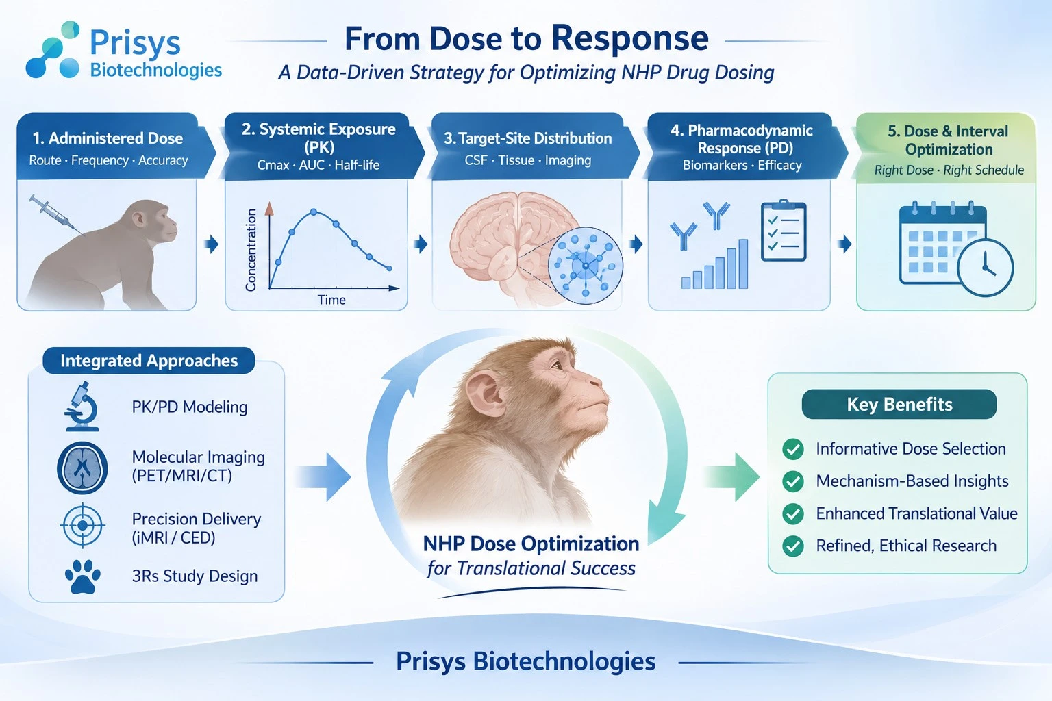
3. Beyond Plasma PK: Understanding Drug Distribution at the Target Site
Systemic exposure describes disposition, but plasma concentration does not establish whether a candidate reaches its pharmacological target.
The gap widens for CNS therapeutics, tissue-targeted biologics, gene therapies, and any modality facing a substantial distribution barrier.
Plasma exposure in CNS programs can run high while brain penetration stays flat. Finding a therapeutic agent in cerebrospinal fluid (CSF) does not establish its concentration in brain parenchyma or in a specific anatomical target either.
CSF and Biological Compartment Assessment
CSF sampling measures exposure inside the central nervous system and shows how the CSF compartment tracks systemic circulation.
Read the result against the drug's physicochemical properties, molecular size, transport mechanisms, and intended site of action.
For biologics and nucleic acid therapeutics, tissue-level evidence is usually needed to show that intact, active material reaches the relevant tissue.
Imaging-Based Biodistribution Assessment
Molecular imaging measures distribution in the living subject.
With an appropriately labeled compound, PET or SPECT gives in vivo distribution and kinetics: tissue uptake, regional distribution, and evidence of target engagement.
The label needs validation first. Tracer stability, radiometabolites, labeling effects, and the gap between imaging signal and intact therapeutic molecule all affect how the images read.
MRI and CT supply the anatomy: disease-related structural change, tissue characteristics, and the anatomical context of drug delivery.
MRI-guided delivery also visualizes anatomical positioning and monitors the infusion as it proceeds.
Pairing systemic PK with imaging-based distribution addresses a question that comes up in most translational programs: does the administered dose produce adequate systemic exposure and achieve sufficient distribution at the intended site of action?
The question matters most for therapies where tissue penetration or regional delivery is the development bottleneck.
4. Precision Delivery: Why Administration Method Matters
The route of administration sets absorption, bioavailability, and distribution, and through them the relationship between dose and response.
Intravenous dosing delivers directly into the systemic circulation, which separates exposure from the absorption phase. Oral dosing brings in gastrointestinal absorption, formulation behavior, and first-pass metabolism. Subcutaneous and intramuscular dosing add absorption profiles that shift with the molecule and the formulation.
For biologics, intravenous versus subcutaneous administration changes both peak exposure and the shape of the concentration-time profile. With long-acting formulations, slow absorption from the injection site shapes the apparent terminal elimination phase.
NHP studies should use the intended clinical route whenever the science and the technique allow it.
CNS programs face a harder version of the same choice. After systemic dosing, many therapeutic candidates reach specific brain structures poorly.
Direct intracranial delivery and convection-enhanced delivery (CED) reach anatomically defined CNS targets that systemic dosing does not.
In these studies, delivery accuracy, infusion parameters, cannula positioning, and distribution within the target structure all shape the resulting pharmacological response.
Combined with PK and PD measurements, imaging separates insufficient drug exposure from inadequate target-site delivery.
At Prisys, MRI-guided neurosurgical procedures using the ClearPoint navigation system and SmartFlow infusion technology support NHP studies involving targeted CNS delivery and CED. The work covers gene therapies, biologics, nucleic acid therapeutics, and other investigational CNS treatments.
5. Linking PK to PD: Identifying the Exposure Required for Efficacy
Dose optimization aims at an exposure range that produces the intended pharmacological response at an acceptable safety margin, not at the highest exposure an animal tolerates.
Pharmacodynamic evaluation provides the biological context that makes a PK result interpretable.
Depending on the therapeutic mechanism, relevant PD endpoints range across circulating biomarkers, receptor occupancy, target engagement, imaging findings, functional assessments, disease-related physiological changes, and indication-specific efficacy measurements.
PK/PD models describe the exposure-response relationship in several ways.
An Emax model handles a saturable exposure-response curve:
E = E0 + (Emax × C) / (EC50 + C)
Where:
- E represents the observed pharmacological effect.
- E0 represents the baseline effect.
- Emax represents the maximum achievable effect.
- C represents the relevant drug concentration.
- EC50 represents the concentration associated with 50% of the maximum effect.
When the response lags, an indirect-response model or an effect-compartment approach fits better. Both describe the case where plasma concentration changes quickly and the biological response develops slowly.
The concentration in the model should come from the pharmacologically relevant compartment whenever the data allow it. Plasma works as a surrogate for some modalities and fails for drugs whose activity depends on tissue penetration or local target engagement.
A CNS candidate often shows a clean relationship between plasma exposure and systemic biomarkers and a weaker one with neurological functional outcomes. Reading CSF, imaging, and functional PD endpoints together gives a better account of that split.
This integrated approach also locates the cause of an apparent lack of efficacy: inadequate systemic exposure, insufficient target-site distribution, incomplete target engagement, or a pharmacological mechanism that does not translate.
6. Optimizing the First Dose and Dosing Interval
Dose selection depends on how much exposure is needed and how long it must last.
Set the first dose from the available pharmacology, preclinical PK data, the relevant exposure-response relationship, and the anticipated safety profile. For biologics and other modalities with complex disposition characteristics, target-mediated clearance and potential immunogenicity shift the expected exposure profile as well.
Set the dosing interval from the duration of pharmacological activity, not from plasma half-life alone.
Two candidates with similar plasma half-lives can differ widely in how long their activity lasts, through differences in target binding, tissue retention, receptor occupancy, or downstream biological effects. A drug can also remain measurable in plasma long after its activity has declined.
Repeated-dose PK/PD studies show whether the selected interval maintains the desired exposure or biological response. Trough concentrations, accumulation ratios, biomarker changes, and functional endpoints each contribute to that assessment.
For long-acting biologics, sustained exposure and target engagement carry most of the weight. For CNS therapeutics delivered through CED, local distribution and persistence within the target region say more than systemic plasma exposure alone.
The schedule that holds up is the one the combined PK, PD, distribution, and safety evidence supports.
7. Improving NHP Study Design Through the 3Rs
Dose optimization and study refinement run together.
A poorly designed study can use more animals than it needs and still produce too little information for a reliable dose decision. Cutting group sizes without checking statistical power breaks the PK/PD interpretation instead.
The 3Rs principles (Replacement, Reduction, and Refinement) belong in the experimental design from the start.
Several design choices raise the scientific value of each NHP study.
Longitudinal assessment takes repeated PK, imaging, biomarker, and functional measurements in the same animals where the science allows it. Within-animal change over time becomes visible, and the study depends less on separate cross-sectional cohorts.
Integrated endpoint selection keeps exposure, imaging, and pharmacological response in one coordinated design instead of three separate experiments.
Statistical planning sets sample size from the primary research objective, the expected effect size, biological variability, and the planned analysis. Randomization, blinding, and appropriate control groups reduce bias and improve the reliability of conclusions.
Where the objectives support it, sequential dose evaluation or an adaptive approach avoids unnecessary dose groups.
Animal reduction should not become a numerical target in isolation. The aim is the smallest number of animals that still yields scientifically interpretable and adequately supported conclusions.
8. Integrating PK/PD and Imaging in NHP Drug Development
Newer therapeutic modalities have raised the requirements for preclinical dose optimization.
For a conventional small molecule, systemic PK and exposure-response analysis cover much of what dose selection needs. Antibodies, nucleic acid therapeutics, gene therapies, and CNS-targeted treatments also require evidence on tissue distribution, target engagement, and biological persistence.
That requirement pulls pharmacology, bioanalytical measurements, imaging, and specialized delivery procedures into one workflow.
Prisys Biotechnologies focuses on non-human primate pharmacology and translational drug evaluation, integrating disease models, PK/PD study execution, clinical imaging, and specialized surgical capabilities where the research objectives call for them.
Its imaging infrastructure includes MRI, CT, PET-CT, and DSA, supporting anatomical assessment and selected in vivo imaging applications. For CNS studies that need anatomically precise delivery, MRI-guided neurosurgical procedures and CED examine delivery accuracy and target-region distribution.
Run inside an appropriately designed NHP study, these approaches measure several dimensions of drug performance at once: how much drug enters systemic circulation, how it distributes within relevant biological compartments, whether it reaches the intended target, and whether the resulting exposure produces a measurable pharmacological response.
Designing More Informative NHP Dosing Studies
Dose optimization in NHP pharmacology is an exposure-response problem, and the mg/kg calculation covers only the first part of it.
A scientifically meaningful dosing strategy establishes the relationship between administered dose, systemic PK, target-site distribution, pharmacodynamic activity, and safety. The relative importance of each component depends on the therapeutic modality, mechanism of action, intended clinical route, and disease model.
For translational drug development, serial PK sampling combined with relevant biological endpoints and imaging-based distribution assessment gives a fuller picture of drug performance. Sound statistical design and longitudinal assessment then raise the information obtained from each NHP study.
At Prisys, these considerations form part of the broader approach to NHP pharmacology and translational research, particularly for therapeutic candidates that require specialized delivery, tissue-level assessment, or integrated PK/PD evaluation.
The question that decides a study design is whether the selected dose delivers adequate exposure to the right biological target for the required duration.
Frequently asked questions
Q: How is the optimal drug dose determined in NHP studies?
A: The optimal dose is determined by integrating PK exposure, pharmacodynamic response, safety findings, and the intended therapeutic target. Parameters such as Cmax, AUC, trough concentration, and exposure-response relationships help establish an appropriate dose range and dosing interval.
Q: Why is plasma PK insufficient for some NHP drug development studies?
A: Plasma PK describes systemic exposure but does not necessarily reflect drug concentration at the intended target tissue. This matters for CNS therapeutics, biologics, gene therapies, and tissue-targeted treatments. CSF sampling, tissue analysis, and molecular imaging provide complementary evidence of drug distribution and target engagement.
Q: How can imaging improve NHP PK/PD studies?
A: PET and SPECT support the investigation of appropriately labeled compounds in vivo. MRI and CT provide anatomical information and support the evaluation of disease progression and delivery procedures. Read with PK and PD measurements, imaging connects systemic exposure, target-site distribution, and pharmacological response.
Q: How should dosing intervals be optimized for biologics?
A: Dosing intervals should be based on the relationship between systemic exposure and the duration of pharmacological activity. Relevant considerations include elimination half-life, target-mediated drug disposition, target engagement, accumulation, immunogenicity, and the persistence of downstream biological effects.
Q: How can the 3Rs principles be incorporated into NHP PK/PD study design?
A: The 3Rs can be incorporated through appropriate sample-size calculations, longitudinal measurements, integrated PK/PD and imaging endpoints, and efficient experimental designs. The objective is to obtain scientifically reliable information while avoiding unnecessary animal use or underpowered studies.
References and Further Reading
- NC3Rs. Key Elements of a Well-Designed Experiment. https://nc3rs.org.uk/3rs-resources/key-elements-well-designed-experiment
- NC3Rs. Minimising Non-Human Primate Use in Drug Development. https://nc3rs.org.uk/3rs-resources/minimising-NHP-use
- National Institutes of Health, Clinical Center. Drug Development: Nonclinical Study Design and Exposure-Response Considerations. https://clinical.nih.gov/manage-clinical-trials/regulatory-toolbox/investigational-new-drugs/drug-development
- FDA. Product Development Under the Animal Rule: Guidance for Industry.
- ICH M3(R2). Nonclinical Safety Studies for the Conduct of Human Clinical Trials and Marketing Authorization for Pharmaceuticals.