Introduction: The Challenge of Translating Musculoskeletal Therapies
Rheumatoid arthritis (RA) and osteoarthritis (OA) bring inflammation, cartilage degeneration, bone remodeling and loss of joint function into the same clinical picture, and each process feeds the others. Understanding of the underlying mechanisms has advanced considerably. Converting that understanding into clinically meaningful outcomes has been slower.
Part of the difficulty is that no single biological or structural endpoint describes a musculoskeletal disease. A fall in inflammatory cytokines is a poor proxy for cartilage preservation. Less synovial inflammation does not by itself restore joint mobility. A change in bone structure says little about whether the animal moves better.
Preclinical programs therefore need to measure several dimensions at once: molecular pharmacology, structural imaging, and function.
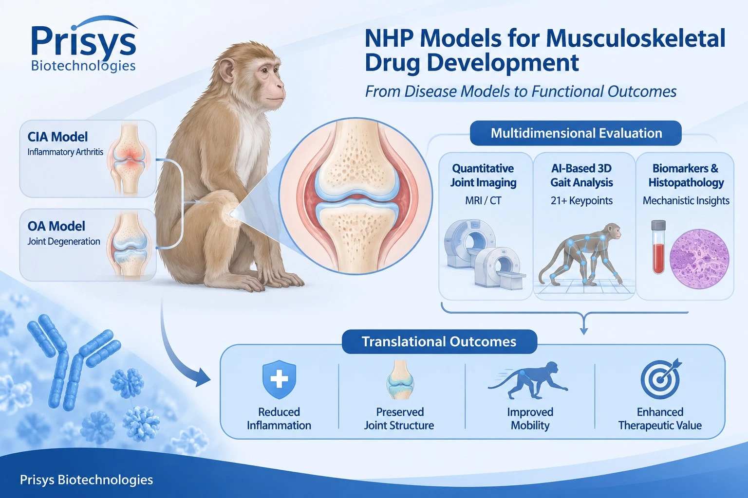
Non-human primate (NHP) models serve as the translational platform for selected musculoskeletal programs. Joint organization, limb biomechanics and immune regulation in macaques sit closer to human joint structure and movement than in conventional laboratory species, and the larger joint dimensions leave room for clinical-grade imaging.
Model choice is only one part of the design. MRI and CT follow joint pathology in the same animal across time points, and AI-based three-dimensional (3D) behavioral analysis adds a functional readout that imaging cannot supply.
For sponsors working on biologics, targeted anti-inflammatory agents or tissue-repair candidates, combining those endpoints gives a fuller account of pharmacological activity and of functional benefit.
1. Why Musculoskeletal Drug Development Requires Multidimensional Evaluation
Musculoskeletal disease involves several interconnected pathological processes. In RA, immune-mediated synovial inflammation drives progressive joint damage. In OA, cartilage degeneration, subchondral bone remodeling, synovial inflammation and altered joint biomechanics all push the disease forward.
The mechanisms differ, but the structural consequence is the same: damage and functional loss develop through interactions across several joint tissues.
Rheumatoid Arthritis: Beyond Suppressing Inflammation
RA is a chronic autoimmune disease marked by persistent synovial inflammation, immune-cell infiltration, synovial hyperplasia, and progressive destruction of cartilage and bone.
Tumor necrosis factor-alpha (TNF-α), interleukin-6 (IL-6) and interleukin-1 beta (IL-1β) drive the inflammatory cascade. Activation of synovial fibroblasts and osteoclasts then carries the damage into cartilage and bone.
Biologic therapies against TNF, IL-6 and other immune pathways changed how RA is treated. Drug development still needs to know how pathway inhibition relates to tissue protection and to functional recovery.
Cytokine concentrations and inflammatory biomarkers alone do not answer that. A program needs to show whether treatment reduces synovial inflammation, limits structural deterioration, and preserves joint function.
That question is sharpest for next-generation biologics: monoclonal antibodies, bispecific antibodies and other targeted immunomodulatory therapeutics.
Osteoarthritis: A Whole-Joint Disease
OA is now read as a disorder of the whole joint rather than cartilage wear alone. Its pathology covers progressive cartilage deterioration, subchondral bone change, osteophyte formation, synovial inflammation and alterations in periarticular tissues.
Where RA centers on immune-mediated synovitis, OA sits at the intersection of mechanical stress, tissue degeneration, low-grade inflammation and abnormal joint remodeling.
An OA candidate may therefore carry several therapeutic objectives at once. Some compounds aim at pain and inflammation, others at cartilage protection, bone remodeling or tissue repair, and each objective needs its own pharmacodynamic endpoint.
A cartilage-sparing candidate cannot be judged on inflammatory biomarkers alone. A candidate intended to improve joint function needs objective functional measurement on top of structural evaluation.
So the preclinical strategy has to combine structural, inflammatory and functional endpoints.
2. Why Non-Human Primate Models Can Support Musculoskeletal Research
No single species reproduces every aspect of human joint disease. Rodent models still do most of the early work: target validation, mechanistic studies and preliminary screening across many compounds, with short experimental cycles and established induction methods.

Joint anatomy, body weight, locomotor biomechanics, immune regulation and tissue dimensions differ enough in rodents that some findings do not carry over.
NHP models cover part of that gap.
Anatomical and Functional Relevance
Macaques share joint organization, limb biomechanics and patterns of coordinated movement with humans. Those similarities matter most when a therapy is meant to preserve function or restore mobility.
Larger joint dimensions also make room for more demanding imaging. Cartilage, synovial tissue, bone and surrounding structures can be followed with clinical imaging equipment and protocols adapted for animal studies.
Immunological Relevance for Biologic Therapies
Species-specific target compatibility decides whether an NHP study is interpretable at all. Monoclonal antibodies, receptor antagonists, cytokine inhibitors and other protein therapeutics bind with different affinities and show different biological activity across species.
A disease model alone does not establish that an NHP is the right species for a given candidate. Target sequence conservation, receptor expression, binding characteristics and functional activity need to be characterized first.
Once those are in hand, an NHP study yields evidence on pharmacological activity, dose-response and exposure-response relationships, and on how target modulation connects to disease-associated endpoints.
Compatibility With Clinical Imaging and Functional Assessment
NHP work also keeps imaging and functional assessment inside the same experimental program, rather than leaving a terminal histopathology readout as the primary evidence.
Longitudinal imaging tracks disease progression and treatment response in individual animals. Quantitative movement analysis then shows whether changes in joint pathology come with measurable functional change.
For disease-modifying therapies that matters, because structural preservation and functional benefit do not develop on the same schedule.
3. CIA and Osteoarthritis Models: Different Models for Different Therapeutic Questions
Model choice should follow the candidate's biological mechanism and the pharmacodynamic endpoints the study is meant to deliver.
For inflammatory arthritis, collagen-induced arthritis (CIA) provides the disease-modeling framework for immune-mediated joint inflammation. For degenerative joint disease, OA models address a different set: cartilage, subchondral bone and joint function.
Collagen-Induced Arthritis (CIA) Model
CIA rests on an immune response against collagen that produces inflammatory change in the joints. It suits work on autoimmune joint inflammation and gives a framework for testing therapies aimed at inflammatory and immune pathways.

Typical applications:
- Anti-inflammatory biologics and immunomodulatory therapies.
- Cytokine-mediated inflammatory pathways.
- Synovial inflammation and joint swelling.
- Structural joint damage.
- Pharmacodynamic response to treatment.
CIA is most useful when the mechanism of action runs through immune activation, inflammatory cytokines or synovial pathology.
CIA is not a replica of human RA, though. Induction method, inflammatory course and the resulting pathology differ between animals and patients, and endpoint interpretation has to allow for that.
Osteoarthritis (OA) Model
OA models follow progressive joint degeneration and the effect of an intervention on joint structure and function. Unlike inflammatory arthritis models, OA work usually requires reading cartilage integrity, subchondral bone, osteophytes, synovial change and joint mobility in the same study.
Applications:
- Cartilage-protective therapies.
- Anti-inflammatory treatment of OA-associated synovitis.
- Therapies targeting cartilage and extracellular matrix homeostasis.
- Tissue-repair and regenerative strategies.
- Structural and functional change during disease progression.
For a candidate intended to modify progression, longitudinal assessment carries most of the information. Inflammatory activity may shift well before structural improvement becomes detectable, so imaging and functional assessment should be timed to the expected pharmacological mechanism.
Matching the Model to the Therapeutic Mechanism
|
Therapeutic objective
|
Relevant model
|
Key evaluation dimensions
|
|
Suppression of immune-mediated joint inflammation
|
CIA
|
Synovial inflammation, inflammatory biomarkers, joint pathology
|
|
Inhibition of inflammatory cytokine pathways
|
CIA
|
Target engagement, PD biomarkers, imaging
|
|
Protection against cartilage degeneration
|
OA
|
Cartilage integrity, joint structure, histopathology
|
|
Regulation of subchondral bone remodeling
|
OA
|
Bone morphology, CT findings, histopathology
|
|
Improvement of joint mobility
|
CIA or OA, depending on pathology
|
3D movement analysis, gait-related parameters
|
|
Evaluation of tissue-repair therapies
|
OA or an appropriately selected joint-injury model
|
Structural recovery, tissue pathology, functional outcomes
|
Model selection follows the therapeutic hypothesis. The models that happen to be available should not decide the question.
4. Quantitative Joint MRI and CT: Visualizing Structural and Inflammatory Changes
Imaging carries a large share of translational musculoskeletal work because it characterizes joint pathology in vivo and follows change over time.
MRI and CT give complementary information. MRI reads soft tissue and inflammatory change; CT reads mineralized tissue and bone architecture in detail. Used together, they strengthen the interpretation of pharmacological effect in NHP arthritis studies.
MRI for Synovial Inflammation and Cartilage Assessment
MRI resolves synovial tissue, cartilage, bone marrow, ligaments and surrounding soft tissue at high resolution. In inflammatory arthritis it captures synovial thickening, joint effusion and other inflammatory or structural abnormalities. Where a contrast-enhanced protocol is appropriate, synovial enhancement adds information on inflammatory activity and vascular change.
In OA studies, MRI supports assessment of cartilage morphology, joint effusion, synovial abnormality and subchondral bone-associated change.
Depending on the protocol and the analysis, the endpoints include the following:
Synovial thickness, enhancement patterns and joint effusion together describe inflammatory activity.
Cartilage thickness, surface integrity and morphological abnormality are read from optimized sequences and image analysis.
MRI also picks up abnormality in subchondral bone and in the surrounding soft tissue.
Repeated imaging allows within-animal comparison of disease-associated change and of treatment response.
The value of MRI is not the anatomical picture alone. It lies in turning structural change into measurable pharmacodynamic endpoints.
A candidate acting on inflammatory pathways may show reduced synovial enhancement or joint effusion early. A cartilage-protective therapy usually needs a longer observation window before structural differences become visible.
CT for Subchondral Bone and Joint Architecture
CT resolves mineralized structures and reads bone morphology. In OA it complements MRI on subchondral bone change, osteophyte formation and other structural abnormality.
Parameters include:
- Subchondral bone morphology.
- Osteophyte formation and distribution.
- Joint-space-associated structural change, read alongside other imaging modalities.
- Bone surface abnormality.
- Three-dimensional reconstruction of the joint.
CT and MRI are not interchangeable. MRI covers soft tissue and cartilage more broadly, while CT is the better tool for bone.
Modality choice follows the disease mechanism, the target tissue and the primary endpoints of the study.
From Imaging Observations to Quantitative Pharmacodynamic Endpoints
Imaging assessment should be standardized across time points: consistent positioning, acquisition protocols, anatomical coverage, image reconstruction and quantitative analysis.
Segmentation and region-of-interest analysis then characterize change in specific joint structures, and imaging findings are read alongside histopathology and other biological measurements.
At Prisys Biotech, the NHP imaging platform runs a 1.5T MRI system and a 40-slice CT system, supporting MRI- and CT-based assessment across disease areas that include joint and spinal imaging. For musculoskeletal work, that is the imaging base for evaluating joint pathology and structural change in NHP models.
5. AI-Based 3D Gait Analysis: Turning Joint Dysfunction Into Quantitative Data
Imaging describes pathology but not how the joint performs in motion. Structural abnormality can be present without change in every functional parameter, and animals sometimes alter movement before structural deterioration shows on imaging.
That is where objective functional endpoints come in, and gait analysis is the most direct of them: it reads locomotion, joint mobility, posture and coordinated limb movement.
Limitations of Conventional Gait Assessment
Traditional gait evaluation relies on visual observation, manual scoring or selected measurements from two-dimensional video.
Those methods catch obvious abnormality. Quantifying subtle change in joint function is harder. Manual scoring varies with observer experience, and 2D recordings introduce perspective error that makes movement across several anatomical planes difficult to reconstruct.
In NHP studies the problem is larger, because locomotion involves coordinated movement across multiple joints and limbs.
A fuller approach tracks anatomical landmarks continuously and reconstructs movement in three dimensions.
Markerless 3D Motion Tracking and 21 Anatomical Key Points
AI-based markerless 3D behavioral analysis pairs synchronized multi-view video with deep-learning pose estimation. The system needs no reflective markers or wearable sensors. It identifies anatomical key points directly from the video and rebuilds movement in three-dimensional space.
The workflow used at Prisys combines multi-view synchronization with 3D skeleton reconstruction and tracks more than 21 anatomical key points. From those data come movement trajectories, body positions, joint mobility parameters and kinematic characteristics.
For musculoskeletal research, the applications are these:
Walking pattern, movement speed, limb coordination and locomotor trajectory are all available as video-derived kinematic parameters.
Joint mobility follows from change in the movement of the relevant anatomical landmarks, subject to appropriate anatomical calibration and validation.
Posture, weight bearing and body positioning add evidence of functional impairment or of recovery.
Repeated recordings track functional change before and after treatment.
Behavioral recognition and biomechanical measurement are different things. Pose estimation identifies movement patterns and anatomical trajectories, while clinical gait parameters such as joint angles or limb loading need calibration, validation and study-specific analytical methods.
Why 3D Gait Analysis Matters in Arthritis and OA Research
In RA, inflammatory joint change leads to discomfort, reduced mobility, altered limb use and compensatory movement patterns. In OA, cartilage degeneration and structural remodeling gradually alter locomotion and joint mechanics.
AI-based 3D gait analysis turns those functional consequences into data.
An anti-inflammatory treatment may reduce synovial inflammation with locomotor activity improving alongside. A cartilage-protective candidate may show a different profile, with structural preservation and functional change arriving over a longer period.
Read together with MRI and CT, gait data show whether change in joint pathology comes with functional improvement.
Gait is not a substitute for pain assessment. Locomotion responds to pain, inflammation, neurological function, motivation, body condition and environmental conditions at the same time. Baseline assessment and complementary endpoints are what make the readout interpretable.
6. Integrating Imaging and 3D Functional Assessment for Biologic Drug Development
Biologic development in musculoskeletal disease asks for more than inhibition of a single molecular target. Candidates act on inflammatory pathways, immune-cell activity, tissue remodeling and cell interactions inside the joint microenvironment.
Monoclonal antibodies, bispecific antibodies, cytokine inhibitors and other targeted protein therapeutics all sit in that frame. An integrated NHP pharmacology study connects exposure and pharmacological activity to change in disease pathology and in function.
Anti-Inflammatory Biologics
For therapies against TNF-α, IL-6 or other inflammatory pathways, the first question is whether pathway inhibition reaches reduced joint inflammation and preserved joint structure.
Dimensions to measure:
- Drug exposure and pharmacokinetic profile.
- Target engagement and pharmacodynamic biomarkers.
- Inflammatory biomarker change.
- MRI assessment of synovial abnormality.
- Joint swelling and clinical observation.
- Functional outcome from behavioral and locomotor assessment.
Reading these together says more about therapeutic activity than any single measurement.
Cartilage-Protective and Tissue-Repair Therapies
For OA candidates built to protect cartilage or promote repair, the design leans on structural and functional endpoints. MRI monitors cartilage morphology and associated joint abnormality, and CT adds subchondral bone architecture.
AI-based 3D movement analysis shows whether structural change comes with functional improvement.
Histopathology stays in the design. Cartilage integrity, extracellular matrix organization and other microscopic features are not fully characterized by imaging.
Establishing Exposure-Response Relationships
For biologics, PK and PD assessment belongs inside the overall study design. Exposure does not translate directly into pharmacological response when target-mediated disposition, receptor occupancy, tissue penetration or immunogenicity is in play.
An NHP study can therefore combine drug concentration measurements, target-related biomarkers, imaging endpoints and functional outcomes to examine exposure-response.
That design answers several development questions:
- Does the candidate achieve sufficient systemic exposure?
- Is the intended pharmacological pathway modulated?
- Does target modulation correspond to reduced joint inflammation?
- Do structural changes come with functional improvement?
- Do different endpoints show consistent dose-response relationships?
The answers feed candidate selection, dose optimization and clinical development planning.
7. Designing More Informative NHP Musculoskeletal Studies
Study value depends on the design as much as on the model. Musculoskeletal work needs attention to baseline variability, disease progression, imaging reproducibility and the link between structural and functional endpoints.
Model Selection and Baseline Characterization
CIA and OA models are chosen against the intended therapeutic mechanism. Before dosing starts, baseline disease characteristics have to be established, and the selected animals need to show sufficiently consistent disease-associated change.
Baseline locomotor behavior matters for any functional study, because individual animals move differently even without disease.
Endpoint Selection and Timing
Different mechanisms need different observation windows. Anti-inflammatory effect tends to appear early, while cartilage protection and tissue remodeling take longer.
Imaging and behavioral schedules should follow the expected onset and duration of pharmacological activity. A cytokine-inhibition study may prioritize early inflammatory endpoints, while a disease-modifying OA program needs repeated structural and functional assessment over a longer period.
Combining Imaging, Biomarkers, and Functional Endpoints
Three dimensions organize the evaluation.
Biological activity covers drug exposure, target engagement and inflammatory biomarkers. Structural response covers MRI and CT findings, with histopathology where appropriate. Functional response covers clinical observation, locomotor activity and AI-assisted 3D movement analysis.
Interpretation should respect how these endpoints relate. A reduction in synovial inflammation without functional improvement may mean that more observation time is needed, that structural damage is already established, or that the treatment does not address other contributors to joint dysfunction.
An improvement in locomotor behavior with no imaging correlate deserves the same caution, and should be read against the other pharmacodynamic findings.
8. Musculoskeletal Research Capabilities at Prisys Biotech
Prisys Biotech provides NHP pharmacology and translational research services, including disease models and imaging-based evaluation relevant to musculoskeletal drug development.
Established capabilities include collagen-induced arthritis (CIA) and osteoarthritis (OA) NHP models, with joint MRI and CT assessment.
The imaging platform combines clinical imaging equipment with analytical workflows for evaluating disease-associated structural change in NHP studies.
AI-based markerless 3D behavioral analysis runs inside the NHP research workflow through collaboration with specialized technology partners, with synchronized multi-view acquisition, 3D reconstruction and tracking of more than 21 anatomical key points.
For musculoskeletal programs, these technologies connect joint pathology to change in movement and functional behavior.
Depending on study objectives, an integrated program can combine disease modeling, pharmacological evaluation, longitudinal imaging, behavioral analysis and relevant biological endpoints. That combination suits biologic anti-inflammatory agents, immunomodulatory therapies, cartilage-protective candidates and other investigational treatments for joint disease.
Future Perspectives: Toward More Objective Translational Endpoints
Musculoskeletal therapeutic development is moving toward an integrated understanding of disease modification.
In inflammatory arthritis, the question is how to separate transient suppression of inflammatory activity from sustained protection against structural joint damage. In OA, disease-modifying development needs a clearer account of how cartilage integrity, subchondral bone remodeling, synovial change and joint function interact as the disease progresses.
Both point the same way: molecular pharmacology alongside imaging and objective functional measurement.
NHP disease models, selected and justified on scientific grounds, give a setting where those relationships can be examined. MRI and CT supply complementary structural information, and AI-assisted 3D motion tracking adds a functional dimension.
For the next generation of biologics and tissue-repair therapeutics, combining these endpoints builds a more complete pharmacological evidence package before clinical development.
Frequently asked questions
Q: What are the advantages of non-human primate models in musculoskeletal drug development?
A: NHP models evaluate musculoskeletal therapies in a larger biological system with joint anatomy, immune characteristics and locomotor function relevant to human research. They suit selected studies on biologic therapeutics, inflammatory arthritis, joint imaging and functional assessment. Their use should be justified by the therapeutic mechanism and by the translational question the study is asking.
Q: What is the difference between CIA and OA models in NHP research?
A: Collagen-induced arthritis (CIA) is an immune-mediated inflammatory arthritis model for autoimmune joint inflammation and for evaluating immunomodulatory therapies. OA models focus on joint degeneration: cartilage deterioration, subchondral bone remodeling and the functional impairment that follows. The two address different disease mechanisms and are selected against the pharmacological objective.
Q: How can MRI be used to evaluate synovial inflammation in NHP arthritis models?
A: MRI visualizes synovial thickening, joint effusion and inflammatory change in and around the joint. Contrast-enhanced MRI adds information on synovial enhancement and inflammatory activity. Repeated imaging shows change over time and treatment-associated response when acquisition and image analysis are standardized.
Q: Can AI-based 3D gait analysis be used to evaluate osteoarthritis drug efficacy?
A: Yes. Markerless 3D motion tracking quantifies locomotor pattern, movement trajectory, posture and joint mobility parameters, and those measurements identify functional change linked to OA progression or treatment. Specific biomechanical endpoints still need anatomical calibration and validation, and gait data are read with imaging and other pharmacodynamic assessments.
Q: Why are imaging and behavioral endpoints important for biologic drug development?
A: Biologics act on inflammatory pathways, tissue remodeling and joint function through different mechanisms, and exposure plus inflammatory biomarkers do not capture therapeutic benefit on their own. PK/PD measurement combined with MRI, CT, histopathology and quantitative behavioral analysis gives complementary evidence on pharmacological activity, structural response and functional outcome.
Q: What types of therapeutics can be evaluated using NHP musculoskeletal models?
A: Monoclonal antibodies, cytokine inhibitors, bispecific antibodies, immunomodulatory agents, cartilage-protective compounds and investigational tissue-repair therapies. Suitability depends on species-specific target compatibility, mechanism of action and the availability of relevant pharmacodynamic endpoints.