

Pharmacokinetic (PK) and pharmacodynamic (PD) studies describe how an investigational therapy behaves in vivo and how exposure relates to biological activity. In conventional preclinical development, PK is characterized through serial measurements of drug concentrations in plasma or other biological matrices, which yield parameters such as maximum concentration, exposure over time, clearance, and half-life.
Systemic exposure, however, does not tell us where a drug reaches within the body, how long it remains in a target tissue, or whether enough exposure is achieved at the site of pharmacological action.
The gap matters for therapies aimed at anatomically restricted or biologically complex tissues, including the central nervous system (CNS), tumors, lymphoid organs, and inflamed tissues. A compound can show adequate plasma exposure while penetrating poorly into the intended target tissue; modest systemic concentrations can also coexist with sustained tissue retention or target engagement.
Imaging-based PK/PD approaches, especially molecular imaging with PET, offer another way to address these questions. With appropriately designed radiolabeled tracers or radiolabeled drug candidates, researchers can follow the spatial distribution and temporal behavior of a compound in vivo. Combined with kinetic modeling, target-specific imaging can also shed light on tissue exposure, receptor binding, and pharmacodynamic response.
In non-human primates (NHPs), imaging-based approaches are especially useful because a single study can integrate systemic PK, tissue distribution, imaging biomarkers, and functional endpoints within a physiologically relevant large-animal model.
From Plasma PK to Tissue-Level Exposure
Traditional plasma PK answers a specific question: how does drug concentration in the systemic circulation change over time?
Typical parameters include Cmax, Tmax, AUC, clearance, and terminal half-life. These measurements remain necessary for dose characterization and exposure-response analysis.
The limitation is that plasma concentration is an indirect measure of exposure at most tissue sites.
A CNS-directed therapy, for example, may produce a measurable plasma concentration while crossing the blood-brain barrier poorly. An antibody or other biologic may show prolonged systemic exposure while distributing unevenly across organs and target tissues.
This points to a real distinction between systemic exposure and site-of-action exposure.
Imaging-based PK addresses part of this spatial dimension by generating serial measurements of radiotracer distribution within anatomically defined regions. PET, for example, can quantify radioactivity within a region of interest over time and, depending on the tracer and experimental design, separate specific target-associated uptake from nonspecific distribution.
The resulting information is not simply another measurement of plasma PK. It is a tissue-level dimension that can be read alongside conventional plasma and tissue analyses.
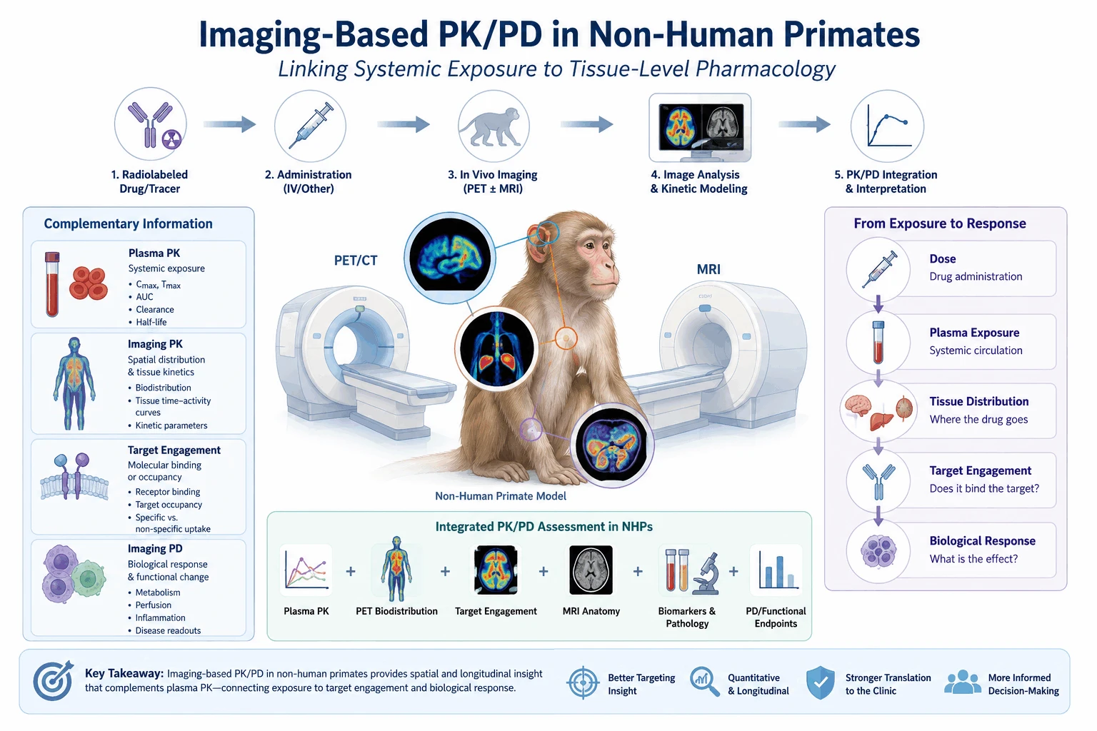
A useful way to think about this is:
Plasma PK → systemic exposure
Imaging PK → spatial distribution and tissue kinetics
Targeted imaging → target-associated binding or engagement
Imaging PD → biological response or functional change
These measurements are complementary rather than interchangeable.
What Is Imaging-Based PK?
Imaging-based PK refers to the use of in vivo imaging to characterize the spatial and temporal behavior of a labeled compound, tracer, or biologically relevant probe.
PET is well suited to this application because radiotracers can be detected at very low concentrations and measured repeatedly over time. Depending on the study design, PET data can yield whole-body biodistribution, tissue-specific uptake, time-activity curves, and kinetic parameters.
Molecular imaging platform materials in translational research group pharmacokinetics, biodistribution, target engagement, and pharmacodynamics as interconnected applications of preclinical imaging. They also stress the quantitative, dynamic nature of PET imaging and its continuity with clinical imaging approaches.
A typical imaging PK workflow may include:
- Radiolabeling or tracer development
- Administration of the labeled compound
- Dynamic or serial PET acquisition
- Anatomical localization using PET/CT or PET/MRI
- Definition of tissue and organ regions of interest
- Generation of time-activity curves
- Kinetic modeling and quantitative analysis
- Integration with plasma PK and, where appropriate, ex vivo tissue measurements
The choice between static and dynamic imaging depends on the scientific question. Static scans can characterize tissue distribution at selected time points, whereas dynamic imaging can show the rate of tissue uptake, retention, and clearance.
For quantitative applications, additional information such as arterial or venous input functions, metabolite analysis, tracer stability, and appropriate compartmental or graphical modeling may be required.
Imaging PK and Conventional PK Provide Different Information
Plasma PK and imaging PK are best understood as two different, overlapping measurements.
| Parameter | Conventional PK | Imaging-Based PK |
|---|---|---|
| Primary measurement | Drug concentration in biological matrix | Radiotracer-derived signal in vivo |
| Main information | Systemic exposure | Spatial distribution and tissue kinetics |
| Sampling | Blood, plasma, tissue | Serial whole-body or regional imaging |
| Spatial information | Limited | Central feature of imaging |
| Target localization | Usually indirect | Can be directly visualized when tracer is target-specific |
| Longitudinal assessment | Requires repeated sampling | Serial non-invasive imaging is possible |
| Quantification | Concentration-based | Activity/kinetic-model-based |
| Key limitation | Limited spatial information | Dependent on tracer behavior and imaging methodology |
This distinction matters when interpreting imaging data.
A PET signal should not automatically be read as the concentration of intact parent drug in a tissue. Radiolabel metabolism, nonspecific binding, blood-pool activity, tracer stability, and partial-volume effects can all influence the measured signal, so appropriate validation is essential.
In well-designed studies, imaging data are integrated with plasma PK, radiometabolite analysis, ex vivo tissue measurements, and pharmacological assays rather than interpreted in isolation.
Imaging Pharmacodynamics: Connecting Tissue Exposure to Biological Activity
Imaging can also contribute to pharmacodynamic assessment.
Traditional PD measurements include circulating biomarkers, cytokines, enzyme activity, receptor occupancy assays, histopathology, and functional behavioral endpoints. They provide evidence of biological activity but may not always show where the response occurs.
Imaging pharmacodynamics, or imaging-based PD, uses molecular or physiological imaging biomarkers to characterize changes associated with drug action in vivo.
Depending on the therapeutic mechanism, imaging PD may include:
- receptor occupancy or target engagement;
- changes in molecular pathway activity;
- changes in tissue perfusion or metabolism;
- changes in inflammatory activity;
- disease-associated structural or functional changes;
- longitudinal changes in lesion or organ characteristics.
This yields a translational sequence that links exposure with pharmacological effect:
Dose → Plasma Exposure → Tissue Distribution → Target Engagement → Biological Response
Observing several stages of this sequence in the same animal is especially useful in NHP studies.
PET for Tissue Distribution and Target Engagement
PET is most useful when the investigational molecule or a pharmacologically relevant ligand can be radiolabeled without substantially altering its biological behavior.
Common radionuclides include 18F, 11C, 64Cu, and 89Zr, with isotope selection depending on molecular size, biological half-life, imaging window, and labeling chemistry. The molecular imaging reference materials describe the use of different PET radionuclides for small molecules, peptides, proteins, antibodies, and cells.
For short-lived small-molecule tracers, dynamic PET may be performed over minutes to several hours. For larger biologics such as antibodies, longer-lived radionuclides such as 89Zr may support imaging over several days.
This flexibility matters for NHP drug development because different therapeutic modalities have substantially different distribution and clearance profiles.
Target-specific PET offers information beyond passive biodistribution. If the tracer binds selectively to a receptor, transporter, enzyme, or other molecular target, tissue uptake can be related to target abundance or binding.
Quantitative kinetic approaches can further improve interpretation.
A representative NHP example is a quantitative PET study of the CD4 pool in rhesus macaques. The investigators used 89Zr-labeled anti-CD4 probes and performed static imaging in 18 animals, while a subset underwent dynamic PET with arterial sampling, metabolite evaluation, and kinetic modeling. The study demonstrated specific uptake in lymph nodes and spleen and used quantitative kinetic analysis to estimate CD4 receptor binding potential.
The example illustrates a key principle: imaging can move beyond visual biodistribution toward quantitative assessment of tissue-specific biological interactions.
The Role of MRI in Imaging-Based PK/PD Studies
MRI and PET supply different types of information.
PET is used for molecular and functional information, whereas MRI provides high-resolution anatomical and tissue characterization. Combining the two modalities can improve spatial interpretation of molecular imaging findings.
In NHP research, MRI can support:
- anatomical localization of PET findings;
- volumetric and structural measurements;
- tissue characterization;
- perfusion and diffusion measurements;
- longitudinal assessment of disease progression;
- image-guided delivery and confirmation of local distribution.
Prisys' clinical imaging platform includes MRI, CT, PET-CT, and DSA, with MRI applications including brain perfusion, diffusion-related imaging, and other advanced post-processing approaches.
For CNS drug delivery studies, MRI plays a different role from conventional imaging PK. During MRI-guided delivery procedures, for example, a contrast agent can serve as a surrogate marker to visualize the distribution of an infused therapeutic formulation. This shows the spatial coverage of the infusion rather than directly measuring the concentration of the therapeutic molecule itself.
This distinction matters when designing and interpreting imaging-based studies.
Why NHPs Are Relevant for Imaging-Based PK/PD
Non-human primates are a useful intermediate model for imaging-based translational pharmacology because they allow systemic PK, molecular imaging, anatomical imaging, and functional endpoints to be evaluated within a large-animal system.
This matters most for CNS therapies, biologics, and other modalities where anatomical scale, vascular organization, tissue accessibility, and molecular target distribution can influence clinical translation.
The combination of NHP models with imaging also supports longitudinal assessment. Rather than relying exclusively on terminal tissue collection, researchers can acquire serial imaging data from the same animals at predefined stages of drug exposure or disease progression.
This longitudinal design can reduce dependence on cross-sectional comparisons and give a more direct view of changes over time.
NHP imaging should not be treated as a replacement for conventional PK or tissue analysis, however. The most informative study designs integrate imaging with plasma PK, tissue sampling where appropriate, biomarker analysis, and pharmacological endpoints.
Integrating Imaging PK/PD into Drug Development
Imaging-based PK/PD can contribute to several stages of translational drug development.
During lead optimization, imaging can help compare candidate molecules according to tissue distribution or target localization.
During preclinical proof-of-concept studies, molecular imaging can show that a biological target is present, accessible, and modulated by treatment.
For CNS programs, imaging can help evaluate whether systemic administration results in measurable exposure in relevant brain regions. For targeted biologics, immuno-PET can help characterize tissue distribution and target-associated uptake.
For cell and gene therapy programs, imaging can support tracking or distribution studies when an appropriate labeling strategy is available.
The molecular imaging framework places biodistribution, pharmacokinetics, target engagement, pharmacodynamics, efficacy, and safety within the preclinical imaging workflow, with corresponding applications extending into clinical development such as dose selection, proof of mechanism, patient selection, and response monitoring.
The value of imaging is not that it replaces existing PK/PD measurements, but that it connects measurements that are otherwise hard to relate spatially.
Key Considerations When Designing Imaging PK/PD Studies
Several methodological factors should be considered before interpreting imaging-derived exposure or PD data.
First, the radiolabeled compound should retain pharmacological behavior sufficiently similar to the unlabeled parent compound. Radiolabeling should not substantially alter affinity, molecular stability, or distribution.
Second, tracer metabolism should be characterized. A PET signal may reflect parent compound, radiolabeled metabolites, nonspecific retention, or a combination of these components.
Third, quantitative analysis should be matched to the biological question. SUV can give a practical semi-quantitative measure, but more rigorous kinetic modeling may be required when the objective is to estimate receptor binding or distinguish specific from nonspecific uptake.
Fourth, imaging should ideally be interpreted together with conventional PK and pharmacological data. Plasma concentration, radiotracer stability, ex vivo tissue analysis, receptor assays, and functional biomarkers can provide essential validation.
Finally, anatomical co-registration is especially important in small or heterogeneous structures. PET findings without accurate anatomical localization may be difficult to interpret, particularly in CNS studies.
Prisys Approach to Integrated NHP Imaging Studies
At Prisys, molecular imaging is incorporated into broader NHP translational research rather than treated as an isolated imaging endpoint. The Translational Research Center combines NHP disease models with clinical-equivalent imaging modalities, PK/PD evaluation, biomarker analysis, pathology, and other pharmacological endpoints.
The imaging infrastructure includes PET-CT together with MRI, CT, and DSA, allowing molecular, anatomical, and physiological findings to be evaluated within the same translational research framework.
Depending on the study objective, an integrated design may combine:
Plasma PK + PET biodistribution + target-specific imaging + MRI/CT anatomical assessment + pharmacodynamic biomarkers
This integration matters most when the central development question is not simply whether a drug enters the circulation, but whether it reaches the intended tissue, interacts with the relevant biological target, and produces a measurable pharmacological response.
Future Perspective
The growing use of molecular imaging in NHP research tracks a shift toward spatially resolved, longitudinal pharmacology.
Conventional PK remains essential for defining systemic exposure. Imaging adds a second dimension, showing where the tracer or drug-associated signal is located and how it changes over time. Target-specific molecular imaging can connect tissue distribution with receptor availability or target engagement, while functional imaging offers additional evidence of pharmacodynamic effect.
The direction is not imaging replacing conventional PK/PD, but a more integrated framework in which systemic exposure, tissue distribution, molecular target engagement, and functional response are read together.
For translational drug development, this approach matters most for therapies where the relationship between plasma exposure and site-of-action exposure is hard to establish with conventional sampling alone.
Conclusion
Imaging-based PK/PD provides a spatial and longitudinal dimension that complements conventional pharmacokinetic and pharmacodynamic measurements.
PET can characterize whole-body biodistribution and tissue kinetics, while target-specific tracers can shed light on molecular binding or target engagement. MRI and other anatomical imaging modalities supply the structural context needed to interpret these molecular signals.
In NHP studies, integrating imaging with plasma PK, biomarkers, tissue analysis, and functional endpoints gives a more complete characterization of the exposure–target–response relationship.
The central principle is not that imaging replaces blood-based PK. Rather, plasma PK describes systemic exposure, while imaging can help determine how that exposure translates into tissue distribution and site-specific pharmacology. This distinction is increasingly relevant for CNS therapies, biologics, targeted therapeutics, and other modalities in which tissue exposure cannot be inferred reliably from circulating concentrations alone.
Recommended reading
Molecular Imaging in NHP Models For Translational Drug Development - News - Prisys
Quantitative PET Target Engagement in NHPs For CNS Drugs
PET Biodistribution Imaging in Non-Human Primates
Molecular Imaging Biomarkers: Advancing Translational Research And Drug Development
Why NHP Molecular Imaging Is Critical For Translational PET Studies
What Is Molecular Imaging And Why Is It Important in Drug Development?
NHP Radionuclide Biodistribution Studies: Molecular Imaging For Translational Drug Development
FAQ
Q: What is imaging-based PK?
A: Imaging-based PK uses in vivo imaging, most commonly PET, to characterize the spatial distribution and time-dependent behavior of a radiolabeled drug, tracer, or pharmacologically relevant probe. It complements conventional plasma PK by providing information about tissue distribution and kinetics.
Q: Is imaging PK a replacement for plasma PK?
A: No. Plasma PK and imaging PK provide different types of information. Plasma PK quantifies systemic exposure, whereas imaging provides spatial information about tissue distribution and, with appropriate tracer design and kinetic modeling, target-associated kinetics.
Q: Can PET directly measure drug concentration in tissue?
A: Not necessarily. PET measures radioactive signal rather than automatically measuring intact parent drug concentration. Interpretation depends on radiolabel stability, tracer metabolism, specific and nonspecific binding, imaging resolution, and the quantitative methodology used.
Q: What is imaging pharmacodynamics?
A: Imaging pharmacodynamics uses molecular or physiological imaging biomarkers to assess biological effects associated with treatment. Examples include receptor occupancy, target engagement, changes in metabolism, perfusion, inflammation, or disease-associated functional changes.
Q: Why combine PET with MRI in NHP studies?
A: PET provides molecular and functional information, while MRI provides high-resolution anatomical and tissue characterization. Combining the modalities can improve anatomical localization and interpretation of molecular imaging findings, particularly in CNS studies.


