


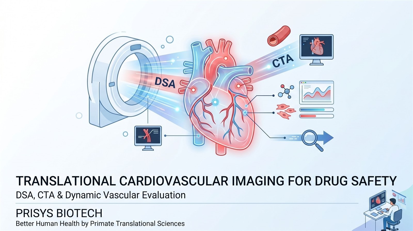
Cardiovascular adverse events remain one of the leading causes of late-stage drug attrition and post-marketing safety concerns. While conventional toxicology and serum biomarker analysis provide valuable information, many vascular complications-including thrombosis, vascular stenosis, embolization, or device-related injury-may develop silently before overt clinical symptoms appear.
With the increasing complexity of cardiovascular therapeutics, biologics, and interventional devices, translational imaging technologies are becoming increasingly important in preclinical safety evaluation. Among these technologies, Digital Subtraction Angiography (DSA) and Computed Tomography Angiography (CTA) have emerged as powerful tools for real-time vascular visualization and longitudinal cardiovascular assessment.
These imaging approaches not only support the evaluation of vascular integrity and hemodynamics, but also provide clinically relevant endpoints that help bridge preclinical findings to human intervention procedures and cardiovascular safety studies.
Why Traditional Cardiovascular Safety Assessments May Be Insufficient
Conventional cardiovascular safety studies often rely on physiological monitoring, blood biomarkers, histopathology, or terminal tissue collection. Although these methods remain essential, they may not fully capture dynamic vascular events occurring during disease progression or therapeutic intervention.
For example, early thrombus formation, partial vessel occlusion, endothelial injury, or altered perfusion may remain undetected until advanced pathology develops. In interventional device studies, procedural complications such as stent malposition, vascular leakage, or restenosis may also require direct imaging confirmation rather than indirect biochemical evaluation.
Furthermore, many cardiovascular therapies-including catheter-based delivery systems, vascular implants, embolization therapies, and targeted biologics-depend heavily on real-time anatomical accuracy and procedural visualization. This creates increasing demand for imaging-guided translational assessment platforms.
What Are DSA and CTA?
Digital Subtraction Angiography (DSA)
DSA is an X-ray–based vascular imaging technology widely used in clinical interventional procedures. By subtracting pre-contrast background images from contrast-enhanced images, DSA enables high-contrast visualization of blood vessels with excellent temporal resolution.
DSA is particularly valuable for:
- Coronary and peripheral vascular imaging
- Interventional catheter guidance
- Stent placement verification
- Vascular occlusion assessment
- Blood flow evaluation
- Embolization procedures
Because DSA provides real-time imaging during intervention, it is commonly used in minimally invasive cardiovascular and neurovascular procedures.
Computed Tomography Angiography (CTA)
CTA combines CT imaging with contrast enhancement to generate high-resolution three-dimensional vascular images. Compared with conventional angiography, CTA offers broader anatomical coverage and detailed structural evaluation.
CTA is frequently applied in:
- Coronary artery visualization
- Aortic disease assessment
- Plaque characterization
- Vascular remodeling analysis
- Stent patency evaluation
- Longitudinal disease monitoring
The ability to reconstruct complex vascular anatomy makes CTA particularly useful for translational cardiovascular research and device evaluation.
Applications of DSA and CTA in Cardiovascular Drug Safety Studies
Evaluation of Thrombosis and Vascular Occlusion
Drug-induced thrombosis remains a major concern in cardiovascular and inflammation-related therapies. DSA and CTA enable direct visualization of vessel patency and thrombus formation, allowing researchers to monitor vascular complications dynamically rather than relying solely on terminal pathology.
In translational models, imaging-based vascular assessment can support:
- Detection of partial or complete vessel occlusion
- Monitoring of thrombus progression
- Quantification of blood flow interruption
- Evaluation of anti-thrombotic therapies
These endpoints may improve the translational relevance of preclinical cardiovascular safety studies.
Interventional Procedure Assessment
Modern cardiovascular research increasingly involves catheterization, vascular access systems, and implantable devices. DSA-guided procedures allow precise visualization during intervention, improving procedural consistency and enabling immediate evaluation of complications.
Applications include:
- Balloon angioplasty
- Stent implantation
- Catheter navigation
- Vascular embolization
- Image-guided drug delivery
- Hybrid surgical procedures
Real-time imaging can help reduce technical variability while improving reproducibility across studies.
Longitudinal Monitoring of Vascular Remodeling
Unlike terminal histological analysis, CTA enables repeated non-invasive imaging throughout the study period. This allows researchers to evaluate disease progression and therapeutic response longitudinally within the same subject.
Longitudinal imaging endpoints may include:
- Vessel diameter changes
- Plaque progression
- Restenosis evaluation
- Calcification assessment
- Perfusion alterations
Such imaging biomarkers are increasingly important for translational cardiovascular drug development.
Translational Advantages of Advanced Imaging in NHP Studies
Non-human primate (NHP) models offer important translational advantages for cardiovascular imaging research due to their anatomical and physiological similarities to humans. Importantly, clinical imaging systems and interventional workflows used in hospitals can often be directly adapted to NHP studies.
This enables more clinically relevant evaluation of:
- Cardiovascular device performance
- Surgical feasibility
- Drug safety signals
- Hemodynamic responses
- Interventional reproducibility
Compared with small animal models, NHP studies also allow the use of clinical-scale catheters, stents, guidewires, and imaging protocols, improving translational continuity from preclinical development to clinical application.
Prisys Biotech's Clinical Imaging Platform
Prisys Biotech has established a Clinical Imaging Platform designed to support translational cardiovascular and interventional research.
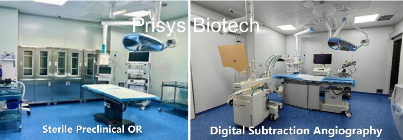
The platform integrates multiple imaging systems, including MRI, CT, PET-CT, and DSA, enabling multimodal evaluation of disease progression, procedural safety, and therapeutic response. According to the platform specifications, the DSA suite supports cardiovascular, neurological, and peripheral vascular intervention procedures, while CTA applications provide detailed arterial visualization and longitudinal vascular assessment.
The imaging infrastructure is supported by clinical-grade operating rooms, hybrid surgical capabilities, and experienced medical imaging teams. The facility also supports minimally invasive procedures, interventional surgery, and image-guided translational studies in non-human primates.
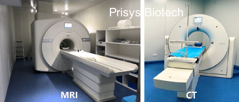
By combining advanced imaging with translational pharmacology and NHP research capabilities, Prisys Biotech aims to support more clinically relevant cardiovascular safety evaluation strategies for emerging therapeutics and medical devices.
Future Perspectives
As cardiovascular therapeutics continue to evolve toward more targeted, device-integrated, and minimally invasive approaches, imaging-based translational evaluation is expected to become increasingly important.
Emerging trends include:
- AI-assisted vascular image analysis
- Quantitative imaging biomarkers
- Real-time perfusion monitoring
- Hybrid surgical imaging workflows
- Image-guided drug delivery systems
- Longitudinal digital endpoints
The integration of DSA, CTA, and advanced translational imaging platforms may help improve early risk identification, optimize procedural safety, and reduce uncertainty during cardiovascular drug development.
FAQ
Q: Why is DSA important in cardiovascular research?
A: DSA provides real-time visualization of blood vessels during interventional procedures, allowing precise assessment of vascular anatomy, blood flow, and procedural outcomes.
Q: What is the difference between CTA and DSA?
A: CTA provides high-resolution three-dimensional vascular imaging using CT technology, while DSA focuses on dynamic real-time angiographic imaging during interventions.
Q: How can imaging improve cardiovascular drug safety evaluation?
A: Imaging technologies help detect vascular abnormalities, thrombosis, stenosis, and procedural complications that may not be fully identified through conventional toxicology endpoints alone.


